

Primary pulmonary Ewing's sarcoma/primitive neuroectodermal tumor in a 67-year-old man
- PMID: 17923745
- PMCID: PMC2694395
- DOI: 10.3346/jkms.2007.22.S.S159
Primary pulmonary Ewing's sarcoma/primitive neuroectodermal tumor in a 67-year-old man
Abstract
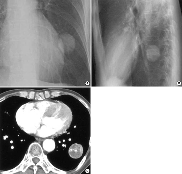
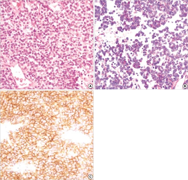
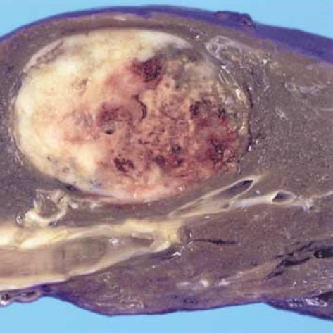
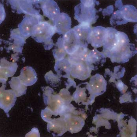
Extraskeletal Ewing's sarcoma (EES) is a branch of neuroectodermal tumor (PNET), which is very rare soft tissue sarcoma. We report a case of EES/PNET arising is the lung of a 67-yr-old man. Computed tomography, bone scintigraphy, and positron emission tomography confirmed the mass to have a primary pulmonary origin. The mass showed positive reactivity in the Periodic Acid Schiff (PAS) stain and MIC-2 immunoreactivity in immunohistochemical stain. Fluorescence in situ hybridization (FISH) was performed, which revealed an EWSR1 (Ewing sarcoma breakpoint region 1) 22q12 rearrangement. The diagnosis was confirmed both pathologically and genetically. The mass lesion was resected, and the patient is currently undergoing chemotherapy.
Figures
References
-
- Tsuji S, Hisaoka M, Morimitsu Y, Hashimoto H, Jimi A, Watanabe J, Eguchi H, Kaneko Y. Peripheral primitive neuroectodermal tumour of the lung: report of two cases. Histopathology. 1998;33:369–374. - PubMed
-
- Imamura F, Funakoshi T, Nakamura S, Mano M, Kodama K, Horai T. Primary primitive neuroectodermal tumor of the lung: report of two cases. Lung Cancer. 2000;27:55–60. - PubMed
-
- Mikami Y, Nakajima M, Hashimoto H, Irei I, Matsushima T, Kawabata S, Manabe T. Primary pulmonary primitive neuroectodermal tumor (PNET). A case report. Pathol Res Pract. 2001;197:113–119. - PubMed
-
- Kahn AG, Avagnina A, Nazar J, Elsner B. Primitive neuroectodermal tumor of the lung. Arch Pathol Lab Med. 2001;125:397–399. - PubMed
-
- Ewing J. Classics in oncology. Diffuse endothelioma of bone. Proceedings of the New York Pathological Society, 1921. CA Cancer J Clin. 1972;22:95–98. - PubMed
Publication types
MeSH terms
Substances
LinkOut - more resources
Full Text Sources
Medical
