

doi: 10.3348/kjr.2007.8.5.429.
The role of PET/CT for evaluating breast cancer
Affiliations
- PMID: 17923786
- PMCID: PMC2626817
- DOI: 10.3348/kjr.2007.8.5.429
Item in Clipboard
The role of PET/CT for evaluating breast cancer
Korean J Radiol.
2007 Sep-Oct.
Abstract
Positron emission tomography combined with computed tomography (PET/CT) has been receiving increasing attention during the recent years for making the diagnosis, for determining the staging and for the follow-up of various malignancies. The PET/CT findings of 58 breast cancer patients (age range: 34-79 years old, mean age: 50 years) were retrospectively compared with the PET or CT scans alone. PET/CT was found to be better than PET or CT alone for detecting small tumors or multiple metastases, for accurately localizing lymph node metastasis and for monitoring the response to chemotherapy in breast cancer patients.
Figures
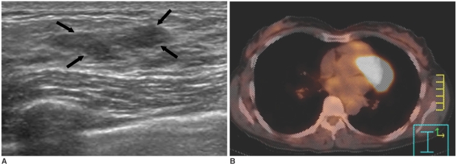
Ductal carcinoma in situ in a 49-year-old woman. A. Sonography shows a 2.5 cm sized hypoechoic mass with an indistinct margin in the left upper breast (arrows). B. The PET/CT image shows no evidence of FDG uptake in the left breast. Surgery revealed ductal carcinoma in situ.
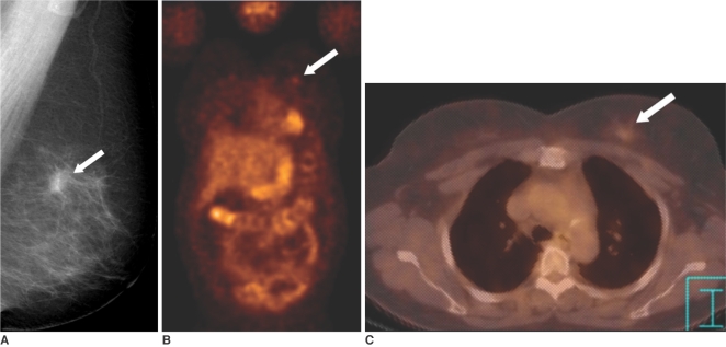
Invasive breast cancer in a 57-year-old woman. A. The mediolateral oblique view of the screening mammogram shows a 1.1 cm sized spiculated mass (arrow) in the left breast. B. The PET image shows faint FDG uptake (SUV = 1.2) (arrow) in the left breast. It is difficult to detect the lesion due to partial volume averaging. C. The PET/CT image shows that the focal uptake (arrow) is localized to the left breast.
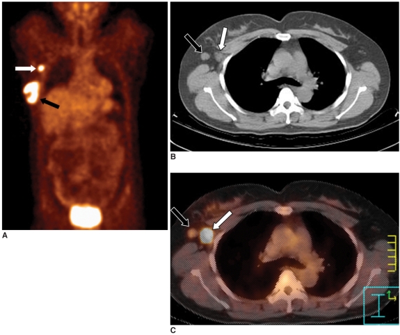
Axillary lymph node metastasis in a 45-year-old woman with a 4 cm invasive ductal carcinoma. A. The PET image shows increased FDG uptake in the right breast (black arrow) and axilla (white arrow). B. The CT image shows two enlarged lymph nodes in the right axilla (arrows). C. The PET/CT image shows accurate localization of the metastatic (white arrow, SUV = 9.9) and reactive (black arrow) lymph nodes. Surgery revealed one metastatic lymph node out of the 21 excised axillary lymph nodes.
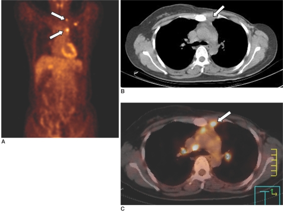
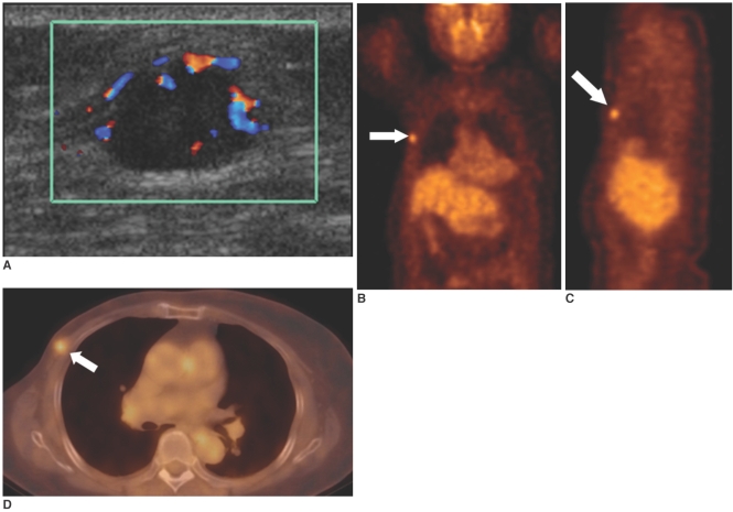
Mediastinal lymph node metastasis in a 41-year-old woman who had undergone left modified radical mastectomy 10 months previously. A. The PET image shows multiple areas of increased uptake (arrows) in the left upper chest. B. The CT image shows a small soft tissue density in the anterior mediastinum (arrow). C. The PET/CT image shows co-registration of the FDG uptake and the soft tissue density in the anterior mediastinum, suggesting internal mammary lymph node metastases (arrow).
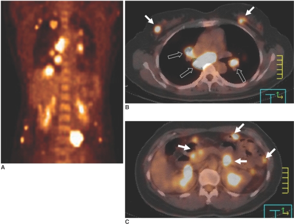
Multiple distant metastases in a 44-year-old woman with bilateral breast cancer. A. The PET image shows multiple areas of FDG uptake in the thorax and abdomen. B, C. The PET/CT images show high uptake in both breasts (white arrows in B), the mediastinal lymph nodes (black arrows in B) and the visceral organs (arrows in C).
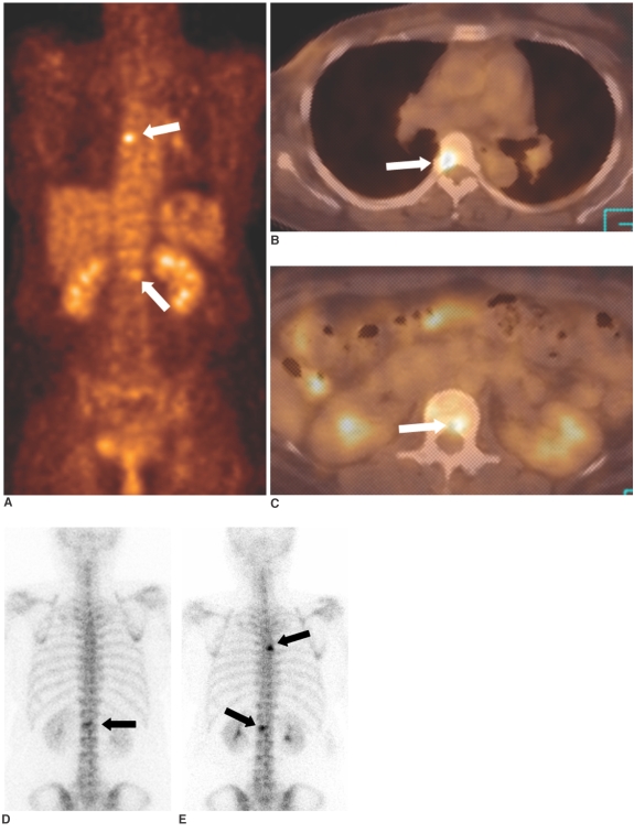
Bone metastases in a 47-year-old woman with breast cancer. A-C. The PET and PET/CT images show increased FDG uptake in the T5 (B) and L1 (C) vertebrae (arrows). Osteolytic changes suggestive of bone metastases are seen on CT. D. The whole body Tc-99m bone scintigram obtained 7 days before the PET scan shows faint uptake in only the L1 vertebra (arrow). T5 metastasis is not visualized. E. The follow-up bone scintigram obtained three months later shows foci of hot uptake in the T5 and L1 vertebrae (arrows). PET/CT was superior to bone scintigram for the early detection of osteolytic breast cancer metastasis.
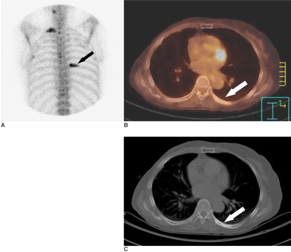
Bone metastases in a 64-year-old woman who had undergone right modified radical mastectomy 36 months previously. A. The bone scintigram shows increased FDG uptake in the right 1st and left 7th ribs (arrow), which is probably due to bony metastases. B. The PET/CT image shows no FDG uptake in the left 7th rib (arrow). C. The CT image shows an osteoblastic bony lesion in the left 7th rib (arrow).
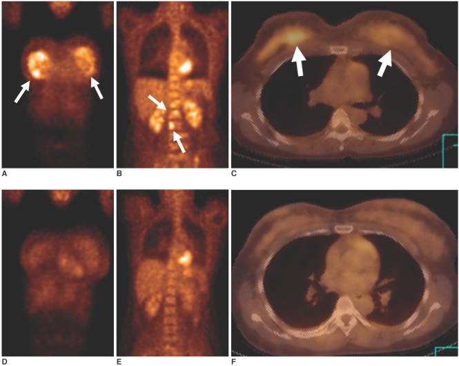
Chemotherapy in a 35-year-old woman with bilateral breast cancer and bone metastases. A-C. The initial PET (A, B) and PET/CT (C) images show strong FDG uptake (arrows) in both breasts and in multiple spinal levels. D-F. The follow-up PET (D, E) and PET/CT (F) images obtained after three cycles of chemotherapy show markedly decreased FDG uptake in both breasts and in multiple spinal levels.
Local recurrence in a 74-year-old woman who had undergone right modified radical mastectomy eight years previously. A. Sonography shows an 1.4 cm oval mass with increased vascularity in the right pectoralis muscle at the mastectomy site. B, C. The PET images show focal high FDG uptake (SUV= 3.3) (arrows) in the right chest wall. D. The PET/CT image shows a focus of high FDG uptake (arrow) localized to the right pectoralis muscle. Accurate localization of the lesion was difficult with using PET alone.
References
-
- Minn H, Soini I. F-18 fluorodeoxyglucose scintigraphy in diagnosis and follow up of treatment in advanced breast cancer. Am J Clin Pathol. 1989;91:535–541. - PubMed
-
- Kubota K, Matsuzawa T, Amemiya A, Kondo M, Fujiwara T, Watanuki S, et al. Imaging of breast cancer with F-18 fluorodeoxyglucose and positron emission tomography. J Comput Assist Tomogr. 1989;13:1097–1098. - PubMed
-
- Wahl RL, Cody RL, Hutchins GD, Mudgett EE. Primary and metastatic breast carcinoma: initial clinical evaluation with PET with the radiolabeled glucose analogue 2-[F-18]-fluoro-2-deoxy-D-glucose. Radiology. 1991;179:765–770. - PubMed
-
- Kostakoglu L, Goldsmith SJ. 18F-FDG PET evaluation of the response to therapy for lymphoma and for breast, lung, and colorectal carcinoma. J Nucl Med. 2003;44:224–239. - PubMed
-
- Beyer T, Townsend DW, Brun T, Kinahan PE, Charron M, Roddy R, et al. A combined PET/CT scanner for clinical oncology. J Nucl Med. 2000;41:1369–1379. - PubMed
