

Case Reports
doi: 10.3348/kjr.2007.8.5.443.
MR imaging in a child with scurvy: a case report
Affiliations
- PMID: 17923788
- PMCID: PMC2626813
- DOI: 10.3348/kjr.2007.8.5.443
Item in Clipboard
Case Reports
MR imaging in a child with scurvy: a case report
Korean J Radiol.
2007 Sep-Oct.
Abstract
Scurvy is very rare disease in industrialized societies. Nevertheless, it still exists in higher risk groups including economically disadvantaged populations with poor nutrition, such as the elderly and chronic alcoholics. The incidence of scurvy in the pediatric population is very low. This study reports a case of scurvy in a 5-year-old girl with cerebral palsy and developmental delay based on MRI findings.
Figures
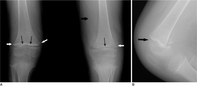
A. Anteroposterior radiograph of both knees shows a thick sclerotic metaphyseal line (thin black arrows) above a widened physis and small beaklike excrescences (white arrows) at the metaphysis in both femora. Soft tissue bulging is noted (thick black arrow). B.Lateral radiograph of the left knee shows a disruption of the alignment of the distal femoral physis (arrow).
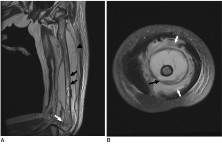
MRI of the left thigh performed at the first hospital day. A. Coronal T2-weighted image shows a diffuse bone marrow signal change (black arrows) of the femur shaft with a large amount of subperiosteal fluid collection (arrowhead) and displacement of the distal epiphysis (white arrow). B.Axial T2-weighted image shows a fluid-fluid level in the subperiosteal fluid collection (black arrow). The surrounding vastus and hamstring muscles also show high intensity signal lesions (white arrows).
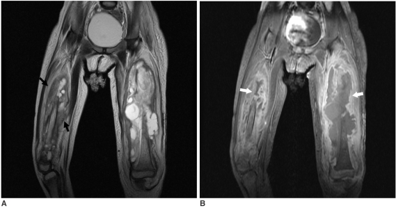
Follow up MR images of both thighs. A. A coronal T2-weighted image shows a larger subperiosteal hematoma at the left femur and a new subperiosteal hematoma (arrows) with signal change of bone marrow of the right femur. B.A coronal contrast enhanced fat suppression T1-weighted image shows moderate enhancement of the periosteum and adjacent soft tissue (white arrows).
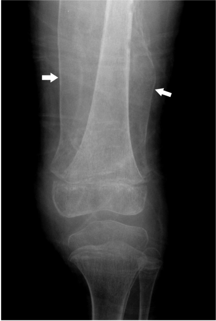
The radiograph performed six weeks after vitamin C supplementation. Large shells of periosteal bone (arrows) are present at the left femur. There was improvement of the metaphyseal sclerotic line and widened physis compared to the initial radiograph.
Similar articles
-
An orange a day keeps the doctor away: scurvy in the year 2000.Pediatrics. 2001 Sep;108(3):E55. doi: 10.1542/peds.108.3.e55. Pediatrics. 2001. PMID: 11533373
-
[Haemorrhages due to vitamin C deficiency. Scurvy in the 21st century].Ned Tijdschr Geneeskd. 2010;154:A1638. Ned Tijdschr Geneeskd. 2010. PMID: 20699018 Dutch.
-
Scurvy presenting with limp and weakness: a case report.BMC Pediatr. 2019 Jul 6;19(1):228. doi: 10.1186/s12887-019-1605-5. BMC Pediatr. 2019. PMID: 31279337 Free PMC article.
-
MRI findings in pediatric patients with scurvy.Skeletal Radiol. 2015 Feb;44(2):291-7. doi: 10.1007/s00256-014-1962-y. Epub 2014 Aug 12. Skeletal Radiol. 2015. PMID: 25109378 Review.
-
An apple a day keeps the doctor away: pediatric scurvy case report and mini review.Childs Nerv Syst. 2024 Sep;40(9):2941-2945. doi: 10.1007/s00381-024-06454-0. Epub 2024 May 16. Childs Nerv Syst. 2024. PMID: 38753002 Review.
Cited by
-
Osteoskeletal manifestations of scurvy: MRI and ultrasound findings.Skeletal Radiol. 2015 Aug;44(8):1161-4. doi: 10.1007/s00256-014-2093-1. Epub 2015 Jan 18. Skeletal Radiol. 2015. PMID: 25597047
-
Lessons learned from "the great mimicker disease": A retrospective study of 18 patients with scurvy.J Child Orthop. 2023 Nov 16;17(6):618-625. doi: 10.1177/18632521231213150. eCollection 2023 Dec. J Child Orthop. 2023. PMID: 38050589 Free PMC article.
-
Fever in Children: Pearls and Pitfalls.Children (Basel). 2017 Sep 1;4(9):81. doi: 10.3390/children4090081. Children (Basel). 2017. PMID: 28862659 Free PMC article. Review.
-
Pediatric scurvy case report: a novel presentation with deep vein thrombosis secondary to large bilateral spontaneous iliac subperiosteal hematomas.BMC Pediatr. 2024 Feb 16;24(1):126. doi: 10.1186/s12887-024-04579-4. BMC Pediatr. 2024. PMID: 38365603 Free PMC article.
-
Magnetic resonance imaging appearance of scurvy with gelatinous bone marrow transformation.Skeletal Radiol. 2012 Mar;41(3):357-60. doi: 10.1007/s00256-011-1350-9. Epub 2012 Jan 6. Skeletal Radiol. 2012. PMID: 22223127
References
-
- Olmedo JM, Yiannias JA, Windgassen EB, Gornet MK. Scurvy: a disease almost forgotten. Int J Dermatol. 2006;45:909–913. - PubMed
-
- Fain O. Musculoskeletal manifestations of scurvy. Joint Bone Spine. 2005;72:124–128. - PubMed
-
- Weinstein M, Babyn P, Zlotkin S. An orange a day keeps the doctor away: scurvy in the year 2000. Pediatrics. 2001;108:E55. - PubMed
-
- Halligan TJ, Russel NG, Dunn WJ, Caldroney SJ, Skelton TB. Identification and treatment of scurvy: a case report. Oral Surg, Oral Med, Oral Path, Oral Radiol, and Endod. 2005;100:688–692. - PubMed
-
- Pimentel L. Scurvy: historical review and current diagnostic approach. Am J Emerg Med. 2003;21:328–332. - PubMed
Publication types
MeSH terms
Substances
LinkOut - more resources
Full Text Sources
Medical
