. 2008 Jan;16(1):131-7.
doi: 10.1038/sj.mt.6300332. Epub 2007 Oct 9.

Efficient in vivo targeting of epidermal stem cells by early gestational intraamniotic injection of lentiviral vector driven by the keratin 5 promoter

Affiliations

Efficient in vivo targeting of epidermal stem cells by early gestational intraamniotic injection of lentiviral vector driven by the keratin 5 promoter

Masayuki Endo et al. Mol Ther. 2008 Jan.

Abstract

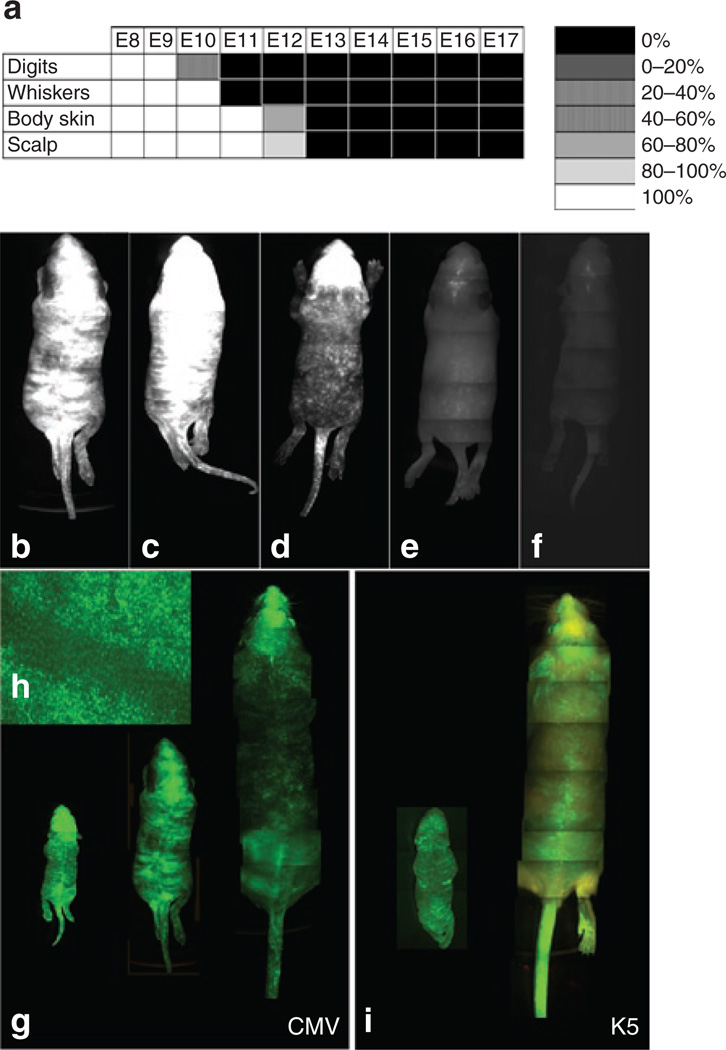
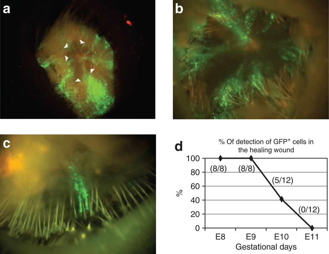
At the present time, no efficient in vivo method for gene transfer to skin stem cells exists. In this study, we hypothesized that early in gestation, specific epidermal stem cell populations may be accessible for gene transfer. To test this hypothesis, we injected lentiviral vectors encoding the green fluorescence protein marker gene driven by either the cytomegalovirus promoter or the keratin 5 (K5) promoter into the murine amniotic space at early developmental stages between embryonic days 8 and 12. This resulted in sustained green fluorescent protein (GFP) expression in both basal epidermal stem cells and bulge cells in the hair follicles of the skin. Transduction of stem cell populations was dependent on the developmental stage, and confirmed by the prolonged duration of GFP expression in all skin elements into adulthood. In addition, transduced stem cell populations responded to regenerative signals after wounding and actively participated in wound healing. Finally, we quantified the fraction of epidermal stem cells transduced, and the distribution of transduction related to the promoters utilized, confirming improved efficiency with the K5 promoter. This simple approach has possible biological applications in our study of gene functions in skin, and perhaps future clinical applications for treatment of skin based disorders.


Figures

Figure 1
Figure 1. The distribution and duration of transgene expression after intraamniotic gene transfer (IAGT) is dependent upon gestational age
(a) Summary of the distribution and frequency of green fluorescent protein (GFP) expression in the skin of mice undergoing IAGT at different gestational time points. IAGT at early gestational ages achieved more widespread expression in a higher number of animals. After E12, no skin GFP expression could be detected by stereoscopic fluorescent microscopy. (b–f) Representative fluorescent stereomicroscopic images of total body GFP expression, 7 days after birth in mice undergoing IAGT from E8 through E12. Mice undergoing IAGT at b E8 and c E9 had diffuse expression with a band-like or striped pattern. Mice undergoing IAGT at d E10 also showed diffuse GFP expression, but in a stippled pattern over the trunk. Mice undergoing IAGT at e E11 and f E12 expressed GFP only on the scalp region with minimal expression on the trunk. (g) GFP expression in mice undergoing IAGT at E8 and E9 maintained GFP expression in a band-like pattern for over 6 months. Representative example of sequential images of a single mouse that underwent IAGT with the lentiviral cytomegalovirus vector at E8 imaged on postnatal days 1 (P1), P7 and P180 (from left to right). (h) A high magnification image of the truncal skin of the same mouse at P180. Note the band-like or striped pattern of fluorescence. (i) Whole body images demonstrating a similar pattern of transduction in P0 and p28 mice that underwent IAGT with the lentiviral-keratin 5 (K5) vector. Fluorescence is obscured somewhat in adult animals by hair growth.
Figure 2
Figure 2. Transduced stem cells participate in the early adult wound healing response
Fluorescence stereomicroscopic image of representative excised wounds of 8-week-old mice that underwent intraamniotic gene transfer (IAGT) at E9, 1week after injury. (a) The punch biopsy wound can be seen within the excised piece of skin. The wound margin is marked by the arrowheads. GFP-positive cells can be seen migrating toward the center of the wound from the wound margin. (b) High magnification of the wound and wound margin from a. (c) Migrating green fluorescent protein (GFP)-positive cells can be seen emerging from individual hair follicles. (d) Frequency of detection of GFP-positive cells participating in wound healing in mice undergoing IAGT at gestational days E8–E11. Mice undergoing IAGT at E8 and E9 uniformly had GFP expression detected in their wounds whereas mice undergoing IAGT at E12 had no GFP-positive cells detected in their healing wounds.
Figure 3
Figure 3. Comparison of gene expression between cytomegalovirus (CMV) and keratin 5 (K5) promoters
Panels (a–j) are representative immunofluorescent or immunohistochemical sections of skin demonstrating that all cutaneous epithelial lineages express green fluorescent protein (GFP) after transduction with lentiviral vectors driven by either the CMV or K5 promoters. Lineages include (a,b) epidermis, (c,d) sebaceous glands, (e,f) hair shaft, (g,h) bulge cells in the hair follicle, and (I,j) the hair bulb. The gestational day of injection and the day of harvest for each specimen were; a E8/P2, b E8/P0, c E8/P21, d E8/P28, e E8/P07, f E8/P28, g E8/P30, h E8/P28, i E8/P70, and j E8/P28. The original magnification/scale for each image are: a,b: ×1,000/25 µm; c–f,i,j: ×400/25 µm; g,h: ×200/50 µm. In the epidermis, there is a difference in the pattern of expression between the two promoters. GFP expression is limited to the suprabasalar layers with the CMV promoter, whereas, with the K5 promoter GFP expression is predominantly in the basal layer. (k) Analysis of GFP expression in skin stem cell populations. Flow cytometric analysis during telogen (50 to 60-day-old mouse) of dorsal skin cells of mice undergoing intraamniotic gene transfer at E8 with either the CMV or K5 promoter. Basal layer cells (α6-integrin positive) and bulge stem cells (α6-integrin/CD34+ cells) were analyzed. In the basal layer cells, 1.23 and 48.2% of cells expressed GFP using the CMV or the K5 promoter, respectively. In bulge stem cells, 0.41 and 13.4% of cells expressed GFP using the CMV or K5 promoter respectively. (l) Quantitative polymerase chain reaction analysis of the presence of the EGFP transgene in genomic DNA of sorted GFP+ and GFP− cell populations. DNA from skin of EGFP transgenic mice served as the positive control. The error bars represented relative quantification maximum.

References

    1. Pulkkinen L, Ringpfeil F, Uitto J. Progress in heritable skin diseases: molecular bases and clinical implications. J Am Acad Dermatol. 2002;47:91–104. - PubMed
    1. Uitto J, Pulkkinen L. The genodermatoses: candidate diseases for gene therapy. Hum Gene Ther. 2000;11:2267–2275. - PubMed
    1. Hengge UR, Bardenheuer W. Gene therapy and the skin. Am J Med Genet C Semin Med Genet. 2004;131C:93–100. - PubMed
    1. Woodley DT, Keene DR, Atha T, Huang Y, Ram R, Kasahara N, et al. Intradermal injection of lentiviral vectors corrects regenerated human dystrophic epidermolysis bullosa skin tissue in vivo. Mol Ther. 2004;10:318–326. - PubMed
    1. Mavilio F, Pellegrini G, Ferrari S, Di Nunzio F, Di Iorio E, Recchia A, et al. Correction of junctional epidermolysis bullosa by transplantation of genetically modified epidermal stem cells. Nat Med. 2006;12:1397–1402. - PubMed

Publication types