Review
. 2007 Nov;49(11):873-88.
doi: 10.1007/s00234-007-0300-7. Epub 2007 Oct 9.

Childhood central nervous system leukemia: historical perspectives, current therapy, and acute neurological sequelae

Affiliations
Review

Childhood central nervous system leukemia: historical perspectives, current therapy, and acute neurological sequelae

Fred H Laningham et al. Neuroradiology. 2007 Nov.

Abstract

Introduction: During the past three decades, improvements in the treatment of childhood leukemia have resulted in high cure rates, particularly for acute lymphoblastic leukemia (ALL). Unfortunately, successful therapy has come with a price, as significant morbidity can result from neurological affects which harm the brain and spinal cord. The expectation and hope is that chemotherapy, as a primary means of CNS therapy, will result in acceptable disease control with less CNS morbidity than has been observed with combinations of chemotherapy and radiotherapy over the past several decades.

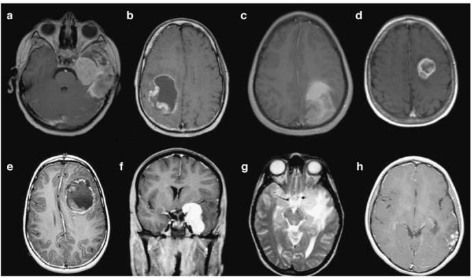
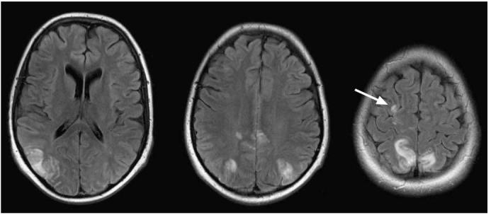
Methods and results: In this review we discuss the poignant, historical aspects of CNS leukemia therapy, outline current methods of systemic and CNS leukemia therapy, and present imaging findings we have encountered in childhood leukemia patients with a variety of acute neurological conditions. A major objective of our research is to understand the neuroimaging correlates of acute and chronic effects of cancer and therapy. Specific features related to CNS leukemia and associated short-term toxicities, both disease- and therapy-related, are emphasized in this review with the specific neuroimaging findings. Specific CNS findings are similarly important when treating acute myelogenous leukemia (AML), and details of leukemic involvement and toxicities are also presented in this entity.

Conclusion: Despite contemporary treatment approaches which favor the use of chemotherapy (including intrathecal therapy) over radiotherapy in the treatment of CNS leukemia, children still occasionally experience morbid neurotoxicity. Standard neuroimaging is sufficient to identify a variety of neurotoxic sequelae in children, and often suggest specific etiologies. Specific neuroimaging findings frequently indicate a need to alter antileukemia therapy. It is important to appreciate that intrathecal and high doses of systemic chemotherapy are not innocuous and are associated with acute, specific, recognizable, and often serious neurological consequences.


Figures

Fig. 1
Fig. 1
CSF involvement in acute lymphoblastic leukemia. The fluid is hypercellular and contains numerous leukemic blasts. This appearance indicates CNS3 status (Wright-Giemsa stain; original magnification ×60, oil immersion) (Courtesy of Mihaela Onciu, MD, St. Jude Children’s Research Hospital)
Fig. 2
Fig. 2
Autopsy specimen with meningeal involvement in acute myeloid leukemia. The meninges are infiltrated by leukemic blasts (arrows), with associated hemorrhage and fibrin deposition (H&E stain; original magnification ×10) (Courtesy of Mihaela Onciu, MD, St. Jude Children’s Research Hospital)
Fig. 3
Fig. 3
Four different patients with leukemia presenting with spine complications at diagnosis. a T2-weighed sagittal MR image shows a thoracic chloroma causing cord compression at T4-7(arrows) of an 11-year-old patient with Ph-positive ALL and lower extremity weakness. Emergent treatment with chemotherapy including steroids resulted in rapid resolution of symptoms. b Sagittal contrast-enhanced T1-weighted image shows tumor in the marrow of the spine in a 6-month-old male presenting with AML. The cord is compressed at T6 (arrow) which caused urinary retention. c Sagittal T1-weighted contrast-enhanced image shows extensive blood products within the epidural and subdural spaces (arrow) in a 5-year old-male with ALL who underwent lumbar puncture at diagnosis while thrombocytopenic at another institution. This improved with time allowing IT therapy to continue. d Sagittal noncontrast-enhanced T1-weighted image of a 10-year-old female who was found to have epidural lipomatosis (arrow) at diagnosis which complicated CNS therapy by impeding CSF acquisition and administration of IT therapy. A ventricular catheter was placed in the brain and successful CNS treatment was given
Fig. 4
Fig. 4
Sacral chloroma (arrows) in an 11-year-old patient with ALL resulting in symptoms of neurogenic bladder. Tumor masses in the confined space of the sacral canal can be particularly symptomatic and more difficult to image
Fig. 5
Fig. 5
An 8-year-old male with a sphenoid bone chloroma near the lateral right orbit which appeared as an enhancing mass on axial T1-weighted contrast-enhanced images. Six months later (right image) the patient had developed a chloroma with intracranial extension into the right cerebellopontine angle. He died shortly after this study from progressive disease including CNS relapse
Fig. 6
Fig. 6
A newborn with congenital ALL presented with seizures. The WBC count was 75×109/l and the platelet count was 3×10⁹/l. CT (left) and MR (center T2-weighted, right T1-weighted) images were obtained 72 h apart. The parietal hemorrhages were thought to be secondary to a combination of hyperleukocytosis and thrombocyto-penia. Fluid-fluid levels with a hematocrit effect are seen within the cystic hemorrhages in the T2-weighted image
Fig. 7
Fig. 7
An 18-year-old male with hyperleukocytosis and thrombocytopenia who complained of left central vision loss and headache and who presented with cerebellar vermis and cerebral hemorrhages detected using noncontrast-enhanced CT. Therapy included leukopheresis. On serial CT examinations, the small hemorrhages and visual acuity improved. The WBC count at diagnosis was 409×109/l and the platelet count was 34×109/l
Fig. 8
Fig. 8
Secondary CNS malignancies in seven children with leukemia, most of whom received cranial radiation therapy for CNS leukemia prevention. a Axial contrast-enhanced T1-weighted image of a 10-year-old treated with BMT for chronic myelogenous leukemia as an infant who developed glioblastoma multiforme (no previous radiation). b Axial contrast-enhanced T1-weighted image of a 24-year-old male treated with craniospinal irradiation as part of therapy for ALL 14 years prior to development of primitive neuroectodermal tumor (PNET). c Axial noncontrast-enhanced T1-weighted image in a 16-year-old male with glioblastoma multiforme developing 13 years after craniospinal irradiation for ALL. d Axial contrast-enhanced T1-weighted image of a 13-year-old male treated with cranial irradiation as part of therapy for ALL. This anaplastic astrocytoma developed 8 years after radiotherapy . e Axial contrast-enhanced T1-weighted image of a 10-year-old male with primitive neuroectodermal tumor 6 years after cranial irradiation for ALL. f, g Coronal contrast-enhanced T1-weighted (f) and axial T2-weighted (g) images of a 20-year-old female treated with cranial irradiation for ALL 17 years earlier developed a meningioma near the left cavernous sinus. h Axial contrast-enhanced T1-weighted image of a 13-year-old female with dysembryoplastic neuroepithelial tumor (DNET) which developed 11 years after cranial irradiation therapy for ALL. This tumor later metastasized
Fig. 9
Fig. 9
MTX myelopathy. This 7-year-old female treated for ALL 2 years earlier, developed an isolated CNS relapse treated with cranial irradiation and five doses of IT MTX. She subsequently developed lower extremity paraparesis with urinary incontinence. Initial MR study of the spine was normal. a Sagittal T2-weighted noncontrast-enhanced image obtained after 2 weeks of persistent symptoms shows T2 hyperintensity along the dorsal aspect of the thoracic spinal cord (thick arrow). b-e Sagittal T2-weighted noncontrast-enhanced (b), sagittal T1-weighted contrast-enhanced (c), axial noncontrast-enhanced T2-weighted (d), and axial contrast-enhanced T1-weighted (e) images after 6 weeks of persistent and stable paraparesis show an increasing T2 signal abnormality dorsally in the cord with involvement of the lateral columns with faint cord enhancement (thin arrows). At the time of this report the child was slowly recovering motor function in the lower extremities and had some improvement in bladder function
Fig. 10
Fig. 10
Left to right: T2-weighted, DW and apparent diffusion coefficient map (ADC map) images. Patient A. 7-year-old female with ALL developed choreoathetosis after IT MTX therapy given 12 days previously. Images show abnormal signal in the caudate head and putamen on the right, presumably correlating with the movement disorder. Patient B. 12-year-old male with ALL treated with one cycle of HD-MTX and IT MTX given 11 days before this MR scan. He was encephalopathic, dysarthric, and had uncontrollable limb movements. Arrows show the abnormalities in each patient
Fig. 11
Fig. 11
A 19-year-old male with a history of severe leukoencephalopathy was being followed with imaging. While asymptomatic, he was found to have cortical vein thrombosis. Imaging (left to right) using 2-D FLASH GRE T1-weighted imaging with subtraction of noncontrast-enhanced and contrast-enhanced T1-weighted sequences helps visualize an isolated cortical vein thrombosis in the patient receiving prednisone and-asparaginase. FLAIR imaging without contrast enhancement also shows the thrombosis
Fig. 12
Fig. 12
DVST in a 6-year-old female with ALL while receivingl-asparaginase, IT therapy, and systemic steroids. The arrows indicate the position of a torcula clot. a, b CT images show density near the torcula (a) and the “empty delta sign” after contrast agent administration (b). c Subtraction of T1-weighted 2-D FLASH images before and after contrast agent administration produces a vascular image depicting the clot as a filling defect. d T2*-weighted GRE image shows blood products within the dural venous sinus. e, f Sagittal GRE T1-weighted MPRAGE images show the clot in the torcula (e) with resolution one week later (f) after treatment with low molecular weight heparin
Fig. 13
Fig. 13
a Infant with AML who developed alpha streptococcus ventriculitis after BMT as part of her AML therapy. Axial non-contrast-enhanced FLAIR image (left) shows edema (arrow) and axial contrast-enhanced T1-weighted image (right) shows enhancement of the ependymal surfaces (arrow). b Patient who died after developing Bacillus cereus bacteremia during ALL therapy. These two sets of non-contrast-enhanced CT images, obtained 50 h apart, show progressive development of cerebral edema. The patient died shortly after the CT scan
Fig. 14
Fig. 14
A 16-year-old male with high-risk ALL underwent BMT and developed seizure. Many small foci of enhancement on T1-weighted contrast-enhanced images of the brain suggest infection. Later, blood cultures grew Stomatococcus mucilaginosus which was treated successfully with antibiotic therapy
Fig. 15
Fig. 15
Posterior reversible encephalopathy syndrome (PRES) in a 15-year-old female patient with ALL during induction therapy. Contrast-enhanced axial FLAIR images after a generalized seizure and hypertension show areas of cortical/subcortical hyperintensity in the distribution of the posterior circulation. All imaging abnormalities resolved in 4 weeks. Watershed distribution hyperintensity is seen more anteriorly in the right hemisphere (arrow)

Similar articles

Cited by

References

    1. Pui CH, Evans WE. Treatment of acute lymphoblastic leukemia. N Engl J Med. 2006;354:166–178. - PubMed
    1. Creutzig U, Zimmermann M, Ritter J, Reinhardt D, Hermann J, Henze G, Jurgens H, Kabisch H, Reiter A, Riehm H, Gadner H, Schellong G. Treatment strategies and long-term results in paediatric patients treated in four consecutive AML-BFM trials. Leukemia. 2005;19:2030–2042. - PubMed
    1. Gibson BE, Wheatley K, Hann IM, Stevens RF, Webb D, Hills RK, De Graaf SS, Harrison CJ. Treatment strategy and long-term results in paediatric patients treated in consecutive UK AML trials. Leukemia. 2005;19:2130–2138. - PubMed
    1. Pui CH, Cheng C, Leung W, Rai SN, Rivera GK, Sandlund JT, Ribeiro RC, Relling MV, Kun LE, Evans WE, Hudson MM. Extended follow-up of long-term survivors of childhood acute lymphoblastic leukemia. N Engl J Med. 2003;349:640–649. - PubMed
    1. Kun LE. Leukemias in children. Pediatric Radiation Oncology. 2007:15–39.

Publication types