. 2007 Nov;81(5):1050-6.
doi: 10.1086/521988. Epub 2007 Sep 18.

Significant correction of disease after postnatal administration of recombinant ectodysplasin A in canine X-linked ectodermal dysplasia

Affiliations

Significant correction of disease after postnatal administration of recombinant ectodysplasin A in canine X-linked ectodermal dysplasia

Margret L Casal et al. Am J Hum Genet. 2007 Nov.

Abstract

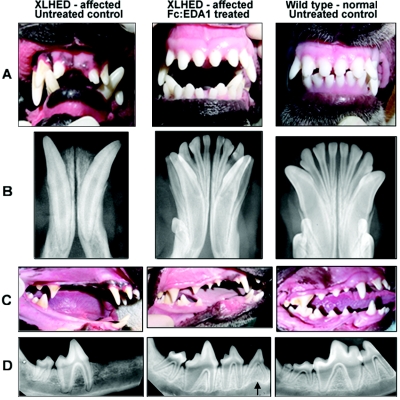
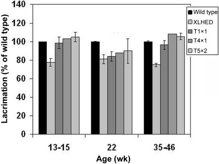
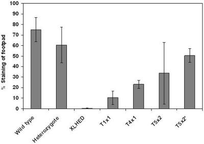
Patients with defective ectodysplasin A (EDA) are affected by X-linked hypohidrotic ectodermal dysplasia (XLHED), a condition characterized by sparse hair, inability to sweat, decreased lacrimation, frequent pulmonary infections, and missing and malformed teeth. The canine model of XLHED was used to study the developmental impact of EDA on secondary dentition, since dogs have an entirely brachyodont, diphyodont dentition similar to that in humans, as opposed to mice, which have only permanent teeth (monophyodont dentition), some of which are very different (aradicular hypsodont) than brachyodont human teeth. Also, clinical signs in humans and dogs with XLHED are virtually identical, whereas several are missing in the murine equivalent. In our model, the genetically missing EDA was compensated for by postnatal intravenous administration of soluble recombinant EDA. Untreated XLHED dogs have an incomplete set of conically shaped teeth similar to those seen in human patients with XLHED. After treatment with EDA, significant normalization of adult teeth was achieved in four of five XLHED dogs. Moreover, treatment restored normal lacrimation and resistance to eye and airway infections and improved sweating ability. These results not only provide proof of concept for a potential treatment of this orphan disease but also demonstrate an essential role of EDA in the development of secondary dentition.


Figures

Figure  1.
Figure 1.
Photographs (A and C) and mandibular dental radiographs (B and D) of wild-type (right), untreated XLHED (left), and treated XLHED (middle) dogs. All incisor and canine teeth were present in the XLHED dog treated with Fc:EDA1 (A and B). The maxillary incisors are normal in shape and number, but both mandibular first incisors and canine teeth were narrowed in diameter compared with their normal counterparts (A). The number of adult premolars is increased in the XLHED dog treated with Fc:EDA1 in comparison with the untreated control XLHED dog, in which most premolars were absent (C). The third premolar (arrow) has the radiographic appearance of a deciduous tooth (D). However, it appears to be an adult tooth on the basis of the clinical appearance of its crown.
Figure  2.
Figure 2.
Mucociliary clearance in XLHED dogs after a single-dose (T1x1, n=3) or multiple-dose (T4x1, n=1; T5x2, n=5; and T5x2*, n=4) treatment with Fc:EDA1. Multiple measurements were obtained for each dog on separate days, to obtain a mean and SD. For illustrative purposes, the data from the dog that did not respond to treatment were removed from the group (T5x2*) and are shown separately (T5x2**).
Figure  3.
Figure 3.
Lacrimal secretions in XLHED dogs after a single-dose (T1x1) or multiple-dose (T4x1 and T5x2) treatment with Fc:EDA1, in percentage of wild type (±SD). The SD is too small to be seen in the normal dogs.
Figure  4.
Figure 4.
Semiquantitative sweat testing in XLHED dogs after a single-dose (T1x1, n=3) or multiple-dose (T4x1, n=1; T5x2, n=5; and T5x2*, n=4) treatment with Fc:EDA1. The ability to sweat was measured on all four feet after oral administration of pilocarpine to induce sweating. For illustrative purposes, the data from the dog that did not respond to treatment were removed from the group (T5x2*).

References

Web Resource

    1. Online Mendelian Inheritance in Man (OMIM), http://www.ncbi.nlm.nih.gov/Omim/ (for XLHED) - PubMed

References

    1. Beahrs JO, Lillington GA, Rosan RC, Russin L, Lindgren JA, Rowley PT (1971) Anhidrotic ectodermal dysplasia: predisposition to bronchial disease. Ann Intern Med 74:92–96 - PubMed
    1. Clarke A (1987) Hypohidrotic ectodermal dysplasia. J Med Genet 24:659–663 - PMC - PubMed
    1. Clarke A, Phillips DI, Brown R, Harper PS (1987) Clinical aspects of X-linked hypohidrotic ectodermal dysplasia. Arch Dis Child 62:989–996 - PMC - PubMed
    1. Kere J, Srivastava AK, Montonen O, Zonana J, Thomas N, Ferguson B, Munoz F, Morgan D, Clarke A, Baybayan P, et al (1996) X-linked anhidrotic (hypohidrotic) ectodermal dysplasia is caused by mutation in a novel transmembrane protein. Nat Genet 13:409–41610.1038/ng0895-409 - DOI - PubMed
    1. Soderholm AL, Kaitila I (1985) Expression of X-linked hypohidrotic ectodermal dysplasia in six males and in their mothers. Clin Genet 28:136–144 - PubMed

Publication types

MeSH terms

LinkOut - more resources