. 2007 Nov;81(5):1098-103.
doi: 10.1086/521953. Epub 2007 Sep 26.

Mutations in TOPORS cause autosomal dominant retinitis pigmentosa with perivascular retinal pigment epithelium atrophy

Affiliations

Mutations in TOPORS cause autosomal dominant retinitis pigmentosa with perivascular retinal pigment epithelium atrophy

Christina F Chakarova et al. Am J Hum Genet. 2007 Nov.

Abstract

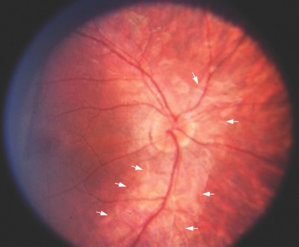
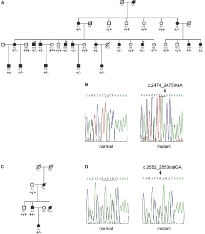
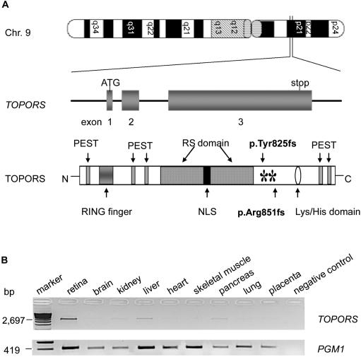
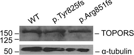
We report mutations in the gene for topoisomerase I-binding RS protein (TOPORS) in patients with autosomal dominant retinitis pigmentosa (adRP) linked to chromosome 9p21.1 (locus RP31). A positional-cloning approach, together with the use of bioinformatics, identified TOPORS (comprising three exons and encoding a protein of 1,045 aa) as the gene responsible for adRP. Mutations that include an insertion and a deletion have been identified in two adRP-affected families--one French Canadian and one German family, respectively. Interestingly, a distinct phenotype is noted at the earlier stages of the disease, with an unusual perivascular cuff of retinal pigment epithelium atrophy, which was found surrounding the superior and inferior arcades in the retina. TOPORS is a RING domain-containing E3 ubiquitin ligase and localizes in the nucleus in speckled loci that are associated with promyelocytic leukemia bodies. The ubiquitous nature of TOPORS expression and a lack of mutant protein in patients are highly suggestive of haploinsufficiency, rather than a dominant negative effect, as the molecular mechanism of the disease and make rescue of the clinical phenotype amenable to somatic gene therapy.


Figures

Figure  1.
Figure 1.
Color photograph of the right eye of a 10-year-old affected child (IV:3). At this age, a very obvious and unusual perivascular “cuff” of atrophy (arrows) around the superior and inferior vascular arcades is visible. The optic disc appears normal in color, although the retinal arterioles are narrow, and there was no pigmentary degeneration at this stage. The cuff of RPE atrophy was found in three other children in this family as well but was not found in the adults of this pedigree. At a later age, this feature apparently disappears.
Figure  2.
Figure 2.
Pedigree structure and sequence analysis of TOPORS mutations. A and C, Canadian and German adRP-affected pedigrees, respectively, used in this study. Affected individuals are shown by blackened symbols, unaffected individuals are identified by unblackened symbols, and deceased individuals are indicated by a slash (/). Asymptomatic individuals are marked by an asterisk (*). Mutation segregation is shown on the pedigree as +/+ (normal) or +/− (affected). B, Electropherogram of the heterozygous mutation, c.2474_2475insA (p.Tyr825fs), in exon 3 of the TOPORS gene found in the Canadian family. D, Electropherogram of the heterozygous mutation found in the German family, c.2552_2553delGA (p.Arg851fs). Both mutations were identified after cloning of the amplified PCR product into pGEMT-Easy vector (Promega).
Figure  3.
Figure 3.
Chromosomal location, domain organization, and expression of TOPORS. A, Schematic representation of exon-intron structure of TOPORS and domain structure of its protein, including the RING-type zinc-finger domain, RS domain, nuclear localization signal (NLS), lysine-histidine domain, PEST region, N-amino-terminal (N), and carboxyl-terminal (C). Both mutations (p.Tyr825fs and p.Arg851fs) are shown with asterisks (*). B, RT-PCR analysis of TOPORS transcript in human tissues: PCR was performed with primers selected from exon 3 (with 28 cycles). A band of 2.69 kb was observed in all the tested QUICK-Clone cDNAs (Clontech) from retina, brain, kidney, liver, heart, skeletal muscle, pancreas, lung, and placenta. A ubiquitously expressed gene, PGM1, was used as a control.
Figure  4.
Figure 4.
TOPORS expression in patient lymphoblastoid cell lines. Protein extracts from cell lines from one unaffected and two affected individuals from the Canadian and German families with p.Tyr825fs and p.Arg851fs mutations, respectively, were analyzed by SDS-PAGE and immunoblotting, with the use of anti-TOPORS antibody (Abnova). α-tubulin was used as a control. WT = wild type.

References

Web Resources

    1. Ensembl Human Genome Browser, http://www.ensembl.org/ (for marker and gene positions)
    1. Online Mendelian Inheritance in Man (OMIM), http://www.ncbi.nlm.nih.gov/Omim/ (for 16 adRP genes, RP31, TOPORS, SLC24A2, and ELAVL2
    1. RetNet, http://www.sph.uth.tmc.edu/Retnet/

References

    1. Rivolta C, Sharon D, DeAngelis MM, Dryja TP (2002) Retinitis pigmentosa and allied diseases: numerous diseases, genes, and inheritance patterns. Hum Mol Genet 11:1219–122710.1093/hmg/11.10.1219 - DOI - PubMed
    1. Hims MM, Diager SP, Inglehearn CF (2003) Retinitis pigmentosa: genes, proteins and prospects. Dev Ophthalmol 37:109–125 - PubMed
    1. Papaioannou M, Chakarova CF, Prescott Q, Waseem N, Theis T, Lopez I, Gill B, Koenekoop RK, Bhattacharya SS (2005) A new locus (RP31) for autosomal dominant retinitis pigmentosa maps to chromosome 9p. Hum Genet 118:501–50310.1007/s00439-005-0063-3 - DOI - PubMed
    1. Sharon D, Yamamoto H, McGee TL, Rabe V, Szerencsei RT, Winkfein RJ, Prinsen CF, Barnes CS, Andreasson S, Fishman GA, et al (2002) Mutated alleles of the rod and cone Na-Ca+K-exchanger genes in patients with retinal diseases. Invest Ophthalmol Vis Sci 43:1971–1979 - PubMed
    1. Antic D, Keene JD (1997) Embryonic lethal abnormal visual RNA-binding proteins involved in growth, differentiation, and posttranscriptional gene expression. Am J Hum Genet 61:273–278 - PMC - PubMed

Publication types

MeSH terms