. 2008 Apr;75(4):632-40.
doi: 10.1002/mrd.20791.

Spermatogenic cell-specific type 1 hexokinase is the predominant hexokinase in sperm

Affiliations

Spermatogenic cell-specific type 1 hexokinase is the predominant hexokinase in sperm

Noriko Nakamura et al. Mol Reprod Dev. 2008 Apr.

Abstract

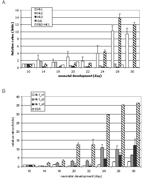
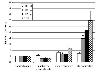
Hexokinase is the first enzyme in the glycolytic pathway and utilizes ATP to convert glucose to glucose-6-phosphate (G6P). We previously identified three variant transcripts of Hk1 that are expressed specifically in spermatogenic cells, have different 5' untranslated regions, and encode a protein (HK1S, spermatogenic cell-specific type 1 hexokinase) in which the porin-binding domain (PBD) of HK1 is replaced by a novel N-terminal spermatogenic cell-specific region (SSR). However, the level of expression of the individual variant transcripts or of the other members of the hexokinase gene family (Hk2, Hk3, and Gck) in spermatogenic cells remains uncertain. We show that Hk1, Hk2, and Hk3 transcripts levels are quite low in spermatocytes and spermatids and Gck transcripts are relatively abundant in spermatids, but that glucokinase (GCK) is not detected in spermatozoa. Using real time RT-PCR (qPCR) with primers specific for each of the three variant forms and RNA from whole testis and isolated germ cells, we found that transcripts for Hk1_v2 and Hk1_v3, but not for Hk1_v1, are relatively high in spermatids. Similar results were seen using spermatogenic cells isolated by laser-capture microdissection (LCM). Immunoblotting studies found that HK1S is abundant in sperm, and immunostaining confirmed that HK1S is located mainly in the principal piece of the sperm flagellum, where other spermatogenic cell-specific glycolytic enzymes have been found. These results strongly suggest that HK1, HK2, HK3, and GCK are unlikely to have a role in glycolysis in sperm and that HK1S encoded by Hk1_v2 and Hk1_v3 serves this role.


Figures

Fig. 1
Fig. 1
The structures of the cDNAs of hexokinase gene-family members. The coding regions of the Hk1 variants and Hk1 Hk2, and Hk3 are similar in length, while that for Gck is approximately half that of the other hexokinase family members. The 5′untranslated regions of Hk1, Hk1_v1, Hk1_v2, and Hk1_3 differ in their lengths and sequences. Pair of facing arrows indicates the positions of the sequence-specific primers used in this study.
Fig. 2
Fig. 2
Expression of hexokinase gene-family members and of Hk1 variants in testis. A: Primers specific for each of the hexokinase gene family members were used to assay by qPCR their transcription in testes of 10 to 30 day-old mice. (Hk1, white bars; Hk2, gray bars; Hk3, black bars; Gck, spotted bars; PBD-Hk1, grey-spotted bars) B: Primers specific for each of the Hk1 variants and for the sequence encoding the SSR were used to assay by qPCR their expression in testes of 10 to 30 day old mice. (Hk1_v1, white bars; Hk1_v2, gray bars; Hk1_v3, black bars; SSR, diagonal line bars) Expression levels were determined as described in Materials and Methods and shown here as ratios (folds) relative to the level on day 10, with that level set at one. Data are expressed as means ± SEM.
Fig. 3
Fig. 3
Expression of Hk1 variants and of Hk1 in isolated spermatogenic cells and of Hk1 transcripts in brain and testis. A: Primers specific for the Hk1 variants (Hk1_v1, Hk1_v2, Hk1_v3) and for Hk1 (primers within the sequence encoding the PBD) were used to assay by qPCR their transcript levels in isolated spermatogenic cells. Expression levels were determined as indicated for Figure 2. (Hk1_v1, white bars; Hk1_v2, gray bars; Hk1_v3, black bars; SSR, spotted bars; Hk1, diagonal line bars; Gck, horizontal line bars; PBD, grey-spotted bars) B: RNA from brain and testis (day 28) were assayed by qPCR using a primer pair within the sequence for the PBD (labeled PBD) and with one primer in the PBD and the other primer in the region common to all Hk1 transcripts (labeled PBD-Hk1). (Brain; white bar; testis, gray bar) Expression levels are shown here as ratios (folds) relative to the level on brain, with that level set at one.
Fig. 4
Fig. 4
Expression of Hk1 variants determined using LCM. RNA from spermatogenic cells isolated using LCM was assayed by qPCR as described in the Methods and Materials. Primers specific for Hk1 variants (Hk1_v1, Hk1_v2, Hk1_v3), Hk1 and SSR used were identical to those used for the studies in Figure 2. Expression levels were determined as indicated for Figure 2. (Hk1_v1, white bars; Hk1_v2, gray bars; Hk1_v3, black bars; SSR, diagonal line bars).
Fig. 5
Fig. 5
Expression of GCK and HK1 in liver, testis and sperm. Protein extracts from liver, testis and sperm were obtained as described in Materials and Methods. Protein (15μg) was loaded in each lane, separated by SDS-PAGE (A and B, right panels) and transferred to membranes. This was repeated at least three times and representative results are shown. The membranes first were probed with antibody to GCK (A, left panel) or to HK1, (B, left panel) and then were stripped and probed with antiserum to the SSR (A and B, middle panels). The antibody to GCK detected a protein only in liver (A, left panel), and the antibody to HK1 detected a protein in sperm (A and B, middle panels). The gels were stained with Coomassie blue to monitor protein loading (A and B, right panel).
Fig. 6
Fig. 6
Immuno-localization of HK1S in mouse testis and sperm. Sections of adult mouse testis were immunostained with antibodies to HK1 (A, B) or to the SSR (C, D). The HK1 antibody reacted strongly with step15–16 spermatids (A, B) and the SSR antibody also reacted with condensing spermatids (C, D). (Bars: 50 μm) Epididymal sperm was stained with antibodies to SSR (E, F) or to HK1 (G, H). The antibody to SSR stained the principal piece region more intensely than the middle piece, but seldom stained head (F). The antibody to HK1 stained the principal piece region and to a lesser degree the middle piece and head (H). Phase micrographs (E, G) show the same sperm that are immuno-stained in the fluorescence micrographs (F, H). (Bars: 8 μm).

Similar articles

Cited by

References

    1. Adams V, Griffin L, Towbin J, Gelb B, Worley K, Macabe ERB. Porin interaction with hexokinase and glycerol kinase: Metabolic microcompartmentation at the outer mitochondrial membrane. Biochem Med Metab Biol. 1991;45:271–291. - PubMed
    1. Arora KK, Fanciulli M, Pedersen PL. Glucose phosphorylation in tumor cells: Cloning, sequencing, and overexpression in active form of a full length cDNA encoding a mitochondrial bindable for hexokinase. J Biol Chem. 1990;265:6481–6488. - PubMed
    1. Beyler SA, Wheat TE, Goldberg E. Binding of antibodies against antigenic domains of murine lactate dehydrogenase-C4 to human and mouse spermatozoa. Biol Reprod. 1985;32:1201–1219. - PubMed
    1. Bluthmann H, Cicurel L, Kuntz GW, Haedenkamp G, Illmensee K. Immunohistochemical localization of mouse testis-specific phosphoglycerate kinase (PGK-2) by monoclonal antibodies. EMBO J. 1982;1:479–484. - PMC - PubMed
    1. Boer PH, Adra CN, Lau YF, McBurney MW. The testis-specific phosphoglycerate kinase gene Pgk-2 is a recruited retroposon. Mol Cell Biol. 1987;7:3107–3112. - PMC - PubMed

Publication types

MeSH terms

LinkOut - more resources