. 2007 Nov;46(5):1404-12.
doi: 10.1002/hep.21849.

Role of alphavbeta6 integrin in acute biliary fibrosis

Affiliations

Role of alphavbeta6 integrin in acute biliary fibrosis

Bruce Wang et al. Hepatology. 2007 Nov.

Abstract

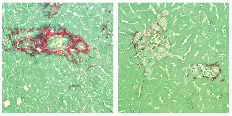
Acute biliary obstruction leads to periductal myofibroblasts and fibrosis, the origin of which is uncertain. Our study provides new information on this question in mice and humans. We show that bile duct obstruction induces a striking increase in cholangiocyte alphavbeta6 integrin and that expression of this integrin is directly linked to fibrogenesis through activation of transforming growth factor beta (TGF-beta). Administration of blocking antibody to alphavbeta6 significantly reduces the extent of acute fibrosis after bile duct ligation. Moreover, in beta6-null mice subjected to the injury, fibrosis is reduced by 50% relative to that seen in wild-type mice, whereas inflammation occurs to the same extent. The data indicate that alphavbeta6, rather than inflammation, is linked to fibrogenesis. It is known that alphavbeta6 binds latent TGF-beta and that binding results in release of active TGFbeta. Consistent with this, intracellular signaling from the TGFbeta receptor is increased after bile duct ligation in wild-type mice but not in beta6(-/-) mice, and a competitive inhibitor of the TGFbeta receptor type II blocks fibrosis to the same extent as antibody to alphavbeta6. In a survey of human liver disease, expression of alphavbeta6 is increased in acute, but not chronic, biliary injury and is localized to cholangiocyte-like cells.

Conclusion: Cholangiocytes respond to acute bile duct obstruction with markedly increased expression of alphavbeta6 integrin, which is closely linked to periductal fibrogenesis. The findings provide a rationale for the use of inhibitors of alphavbeta6 integrin or TGFbeta for down-regulating fibrosis in the setting of acute or ongoing biliary injury.


Conflict of interest statement

Potential conflict of interest: Dr. Violette and Dr. Weinreb own stock in Biogen Idec.

Figures

Fig. 1
Fig. 1
The histological response to selective ligation of the left bile duct (LBDL). Top left: Hematoxylin-eosin (HE) stain showing typical ductal proliferation and mesenchymal expansion in the portal area. Top right: The same area stained for collagen with Sirius red. The nonobstructed lobes from these mice were normal. Below: Hepatocellular injury, as reflected in serum ALT, following acute common (total) bile duct ligation (CBDL) or selective left bile duct ligation (LBDL).
Fig. 2
Fig. 2
Expression of αvβ6 integrin in normal mouse liver (“control”) and at 9, 14, or 16 days after common bile duct ligation. The normal liver lacks detectable αvβ6: the arrowhead indicates a bile ductule. In the liver after bile duct ligation, red fluorescence indicates αvβ6, which is present exclusively in biliary epithelial cells. A pan-cytokeratin antibody (green fluorescence) identifies epithelial cells (hepatocytes and biliary epithelium), whereas a 4′,6-diamidino-2-phenylindole (DAPI) stain (blue) marks nuclei. In the ligated liver, the nuclei surrounding the β6-positive ductules represent injury-associated myofibroblasts (keratin-negative). The appearance is typical. Essentially every bile ductule in all sections examined was strongly positive for αvβ6.
Fig. 3
Fig. 3
Expression of smooth muscle actin (SMA) after bile duct ligation (day 9). SMA-positive cells are in red, and epithelial cells are marked with a pan-cytokeratin antibody (green). On the left is a portal tract of a normal liver. SMA is present in the artery and portal venule; a bile ductule (arrow) is entirely negative. On the right is liver after common bile duct ligation, showing a portion of a medium-sized bile ductule with a large collar of SMA-positive cells. The section shows a relatively large ductule but is representative of ductules of any size. The magnitude of the mesenchymal reaction varied but was uniformly present.
Fig. 4
Fig. 4
Lack of stellate cell migration acutely after bile duct ligation. Mice underwent LBDL. Two weeks later, liver was processed and stained for glial fibrillary acidic protein (GFAP). The section on the left is representative and shows, in the lower portion of the photo, an expanded portal area secondary to biliary obstruction. The stained cells have the typical morphology of stellate cells, which are in their normal position within the lobule and show no tendency to cluster in the portal area. The section on the right is a control, with normal immunoglobulin G in place of the primary antibody (anti-GFAP).
Fig. 5
Fig. 5
Reactive cholangiocytes express smooth muscle actin. The section is from a liver on day 16 after total bile duct ligation, stained with a pancytokeratin antibody (green) and an antibody to smooth muscle actin (SMA, red). The lumen of 3 bile ductules is marked with an asterisk (*). In the ductule with the largest cuff of SMA-positive cells, individual cholangiocytes display red staining in situ (arrows). Of note also, at this stage of the injury, the surrounding SMA-positive cells have an epithelioid shape rather than the spindle shape of typical myofibroblasts, further suggesting that they are cholangiocytes in transition to myofibroblast-like cells. The photomicrograph is representative in that only some cholangiocytes in a ductule appeared to express smooth-muscle actin, and some ductules were entirely negative.
Fig. 6
Fig. 6
Expression of αvβ6 in human liver (see text). The specimen is from a 59-year-old man with a posttransplantation acute biliary obstruction. On the left, the peroxidase stain shows αvβ6, which stains proliferating bile ductules exclusively. On the right is the same sample treated with nonspecific primary antibody. The view is representative.
Fig. 7
Fig. 7
Periductular fibrosis in 6-null or wild-type mice subjected to selective left bile duct ligation (LBDL). The mice were sacrificed 3 weeks after the procedure, and liver sections were stained for collagen using Sirius red. In this representative view, collagen deposition is reduced in the 6-null liver (right panel) while ductule proliferation is increased.
Fig. 8
Fig. 8
Quantitation of collagen deposition in OVA-BIL, β6 null (KO) or wild-type mice. The OVA-BIL mice were studied at 0, 10, and 28 days after adoptive transfer of OVA-specific splenocytes (see Materials and Methods). The β6 null and wild-type mice were subjected to LBDL and evaluated 2 weeks later. Collagen on liver sections was quantified by Sirius red staining and image analysis. Collagen associated with vascular structures was excluded. N = 6 for all groups.
Fig. 9
Fig. 9
Quantitation of periportal granulocytes in wild-type or αvβ6 null mice after selective bile duct ligation. Sections were stained with antibody to the granulocyte antigen Ly-6G as described in Materials and Methods.
Fig. 10
Fig. 10
Immunohistological assessment of signaling from the TGF receptor after bile duct ligation. Wild-type and αvβ6-null mice, respectively, underwent selective bile duct ligation. Six days later, the liver was processed for phospho-Smad2 staining, as described in Materials and Methods. The periportal reaction includes ductule profiles (arrows) as well as an extensive periductal reaction. In liver from wild-type mice, both cholangiocytes and stromal cells express pSmad2 (brown stain), whereas the sections from αvβ6-null mice are negative. The view is representative: essentially all portal areas were positive in the wild-type liver after bile duct obstruction.
Fig. 11
Fig. 11
Effect of blocking antibody to β6 integrin or soluble TGF-β receptor on collagen deposition after bile duct ligation. Mice underwent selective bile duct ligation followed by administration of either a blocking antibody to αvβ6 integrin type II TGF-β soluble receptor, or a control antibody, as described in Materials and Methods. Blocking antibody to αvβ6 integrin and soluble type II TGF-β receptor both significantly reduced collagen deposition compared with control antibody (P = 0.040 and P = 0.036). The amount of reduction by soluble TGF-β receptor is similar to that in β6 knockout mice (n = 7 for anti-β6 group, n = 6 for sTBR and control antibody groups).

References

    1. Roskams T, Desmet V. Ductular reaction and its diagnostic significance. Semin Diagn Pathol. 1998;15:259–269. - PubMed
    1. Alpini G, McGill JM, Larusso NF. The pathobiology of biliary epithelia. Hepatology. 2002;35:1256–1268. - PubMed
    1. Jarnagin WR, Rockey DC, Koteliansky VE, Wang SS, Bissell DM. Expression of variant fibronectins in wound healing: cellular source and biological activity of the EIIIA segment in rat hepatic fibrogenesis. J Cell Biol. 1994;127:2037–2048. - PMC - PubMed
    1. Bissell DM. Chronic liver injury, TGF-beta, and cancer. Exp Mol Med. 2001;33:179–190. - PubMed
    1. Annes JP, Munger JS, Rifkin DB. Making sense of latent TGFbeta activation. J Cell Sci. 2003;116:217–224. - PubMed

Publication types