

Differential long-term neurotoxicity of HIV-1 proteins in the rat hippocampal formation: a design-based stereological study
- PMID: 17924522
- PMCID: PMC3742376
- DOI: 10.1002/hipo.20376
Differential long-term neurotoxicity of HIV-1 proteins in the rat hippocampal formation: a design-based stereological study
Abstract
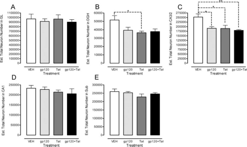
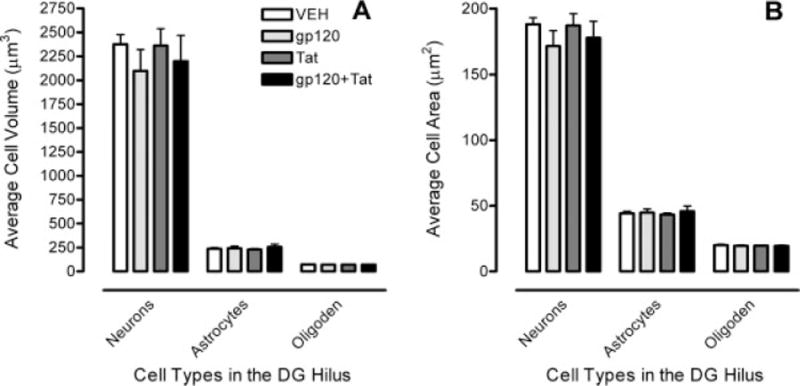
The human immunodeficiency virus type 1 (HIV-1) proteins, gp120 and Tat, are believed to play a role in mediating central nervous system (CNS) pathology in HIV-1 infected patients. Using design-based stereology, we examined the role of neonatal intrahippocampal injections of gp120 and Tat on the adult hippocampus ( approximately 7(1/2) month). Postnatal day (P)1-treated Sprague-Dawley rats were bilaterally injected with vehicle (VEH, 0.5 microl sterile buffer), gp120 (100 ng), Tat (25 microg) or combined gp120 + Tat (100 ng + 25 microg). Using Nissl-stained tissue sections, we quantified total neurons in five subregions of the rat hippocampus [granual layer (GL), hilus of the dentate gyrus (DGH), cornu ammonis fields (CA)2/3, CA1, and subiculum (SUB)], and total glial cells (astrocytes and oligodendrocytes) in two subregions (DGH and SUB). Estimates of cell area and cell volume were taken in the DGH. There was a significant reduction of neuron number in the CA2/3 subfield by Tat and gp120, and a significant reduction in the DGH by Tat only. For glial cells, numbers of astrocytes in the DGH and SUB were increased by the Tat protein, whereas no effects were noted for gp120. Finally, for oligodendrocytes Tat increased cell number in the DGH but not in any other region; gp120 had no detectable effect in any brain region. Estimates of cell area and cell volume of the three different cell types revealed no significant differences between treatments. Collectively, these results suggest differential effects of gp120 and Tat on the estimated total number of neurons, as well as on the number of glial cells.
(c) 2007 Wiley-Liss, Inc.
Figures


Similar articles
-
Neonatal intrahippocampal injection of the HIV-1 proteins gp120 and Tat: differential effects on behavior and the relationship to stereological hippocampal measures.Brain Res. 2008 Sep 26;1232:139-54. doi: 10.1016/j.brainres.2008.07.032. Epub 2008 Jul 17. Brain Res. 2008. PMID: 18674522 Free PMC article.
-
Dose-dependent long-term effects of Tat in the rat hippocampal formation: a design-based stereological study.Hippocampus. 2010 Apr;20(4):469-80. doi: 10.1002/hipo.20648. Hippocampus. 2010. PMID: 19489004 Free PMC article.
-
Neurotoxicity of HIV-1 proteins gp120 and Tat in the rat striatum.Brain Res. 2000 Oct 6;879(1-2):42-9. doi: 10.1016/s0006-8993(00)02725-6. Brain Res. 2000. PMID: 11011004
-
Calcium dysregulation and neuronal apoptosis by the HIV-1 proteins Tat and gp120.J Acquir Immune Defic Syndr. 2002 Oct 1;31 Suppl 2:S55-61. doi: 10.1097/00126334-200210012-00005. J Acquir Immune Defic Syndr. 2002. PMID: 12394783 Review.
-
Brain-derived neurotrophic factor prevents human immunodeficiency virus type 1 protein gp120 neurotoxicity in the rat nigrostriatal system.Ann N Y Acad Sci. 2007 Dec;1122:144-54. doi: 10.1196/annals.1403.010. Ann N Y Acad Sci. 2007. PMID: 18077570 Review.
Cited by
-
Dose-dependent neurocognitive deficits following postnatal day 10 HIV-1 viral protein exposure: Relationship to hippocampal anatomy parameters.Int J Dev Neurosci. 2018 Apr;65:66-82. doi: 10.1016/j.ijdevneu.2017.10.009. Epub 2017 Oct 27. Int J Dev Neurosci. 2018. PMID: 29111178 Free PMC article.
-
HIV-1 Tat and morphine have interactive effects on oligodendrocyte survival and morphology.Glia. 2009 Jan 15;57(2):194-206. doi: 10.1002/glia.20746. Glia. 2009. PMID: 18756534 Free PMC article.
-
In vivo microdialysis in awake, freely moving rats demonstrates HIV-1 Tat-induced alterations in dopamine transmission.Synapse. 2009 Mar;63(3):181-5. doi: 10.1002/syn.20594. Synapse. 2009. PMID: 19086089 Free PMC article.
-
Chronic SSRI treatment reverses HIV-1 protein-mediated synaptodendritic damage.J Neurovirol. 2021 Jun;27(3):403-421. doi: 10.1007/s13365-021-00960-6. Epub 2021 May 18. J Neurovirol. 2021. PMID: 34003469 Free PMC article.
-
Developmental underpinnings of differences in rodent novelty-seeking and emotional reactivity.Eur J Neurosci. 2011 Sep;34(6):994-1005. doi: 10.1111/j.1460-9568.2011.07811.x. Epub 2011 Aug 22. Eur J Neurosci. 2011. PMID: 21864320 Free PMC article.
References
-
- Aksenov MY, Hasselrot U, Bansal AK, Wu G, Nath A, Anderson C, Mactutus CF, Booze RM. Oxidative damage induced by the injection of HIV-1 Tat protein in the rat striatum. Neurosci Lett. 2001;305:5–8. - PubMed
-
- Aksenov MY, Hasselrot U, Wu G, Nath A, Anderson C, Mactutus CF, Booze RM. Temporal relationships between HIV-1 Tat-induced neuronal degeneration, OX-42 immunoreactivity, reactive astrocytosis, and protein oxidation in the rat striatum. Brain Res. 2003;987:1–9. - PubMed
-
- Aksenova MV, Aksenov MY, Mactutus CF, Booze RM. Cell culture models of oxidative stress and injury in the central nervous system. Curr Neurovasc Res. 2005;2:73–89. - PubMed
-
- Aksenov MY, Aksenova MV, Nath A, Ray PD, Mactutus CF, Booze RM. Cocaine-mediated enhancement of Tat toxicity in rat hippocampal cell cultures: The role of oxidative stress and D1 dopamine receptor. Neurotoxicology. 2006a;27:217–228. - PubMed
-
- Aksenova MV, Silvers JM, Aksenov MY, Nath A, Ray PD, Mactutus CF, Booze RM. HIV-1 Tat neurotoxicity in primary cultures of rat midbrain fetal neurons: Changes in dopamine transporter binding and immunoreactivity. Neurosci Lett. 2006b;395:235–239. - PubMed
Publication types
MeSH terms
Substances
Grants and funding
LinkOut - more resources
Full Text Sources
Miscellaneous
