

Stroke induces histamine accumulation and mast cell degranulation in the neonatal rat brain
- PMID: 17924984
- PMCID: PMC8095606
- DOI: 10.1111/j.1750-3639.2007.00092.x
Stroke induces histamine accumulation and mast cell degranulation in the neonatal rat brain
Abstract
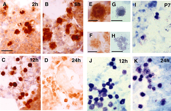
Inflammatory processes are a major cause of hypoxic-ischemic brain damage. The present study focuses on both the cerebral histamine system and mast cells in a model of transient focal ischemia induced by permanent left middle cerebral artery, and homolateral transient common carotid artery occlusion (50 minutes) in the P7 newborn rat. Immunohistochemical analysis revealed that ischemia induces histamine (HA) accumulation in the core of the infarct 6-12 h post-ischemia, and in the penumbra at 24-48 h, although in situ hybridization failed to detect any histidine decarboxylase gene transcripts in these regions. Immunohistochemical co-localization of HA with the MAP2 marker revealed that HA accumulates in neuronal cells before they degenerate, and is accompanied by a very significant increase in the number of mast cells at 12 h and 48 h of reperfusion. In mast cells, histamine immunoreactivity is detected at 2, 6 and 12 h after ischemia, whereas it disappears at 24 h, when a concomitant degranulation of mast cells is observed. Taken together, these data suggest that the recruitment of cerebral mast cells releasing histamine may contribute to ischemia-induced neuronal death in the immature brain.
Figures


Similar articles
-
Stabilizing histamine release in gut mast cells mitigates peripheral and central inflammation after stroke.J Neuroinflammation. 2023 Oct 7;20(1):230. doi: 10.1186/s12974-023-02887-7. J Neuroinflammation. 2023. PMID: 37805585 Free PMC article.
-
Evolution of brain injury after transient middle cerebral artery occlusion in neonatal rats.Stroke. 2000 Jul;31(7):1752-61. doi: 10.1161/01.str.31.7.1752. Stroke. 2000. PMID: 10884483
-
Evaluation of cyclosporine A in a stroke model in the immature rat brain.Exp Neurol. 2011 Jul;230(1):58-66. doi: 10.1016/j.expneurol.2010.06.009. Epub 2010 Jun 16. Exp Neurol. 2011. PMID: 20599982
-
[Development of the research in the field of histamine release].Yakugaku Zasshi. 1994 Mar;114(3):147-59. doi: 10.1248/yakushi1947.114.3_147. Yakugaku Zasshi. 1994. PMID: 7514663 Review. Japanese.
-
Immunohistochemistry in postmortem diagnosis of acute cerebral hypoxia and ischemia: A systematic review.Medicine (Baltimore). 2021 Jun 25;100(25):e26486. doi: 10.1097/MD.0000000000026486. Medicine (Baltimore). 2021. PMID: 34160462 Free PMC article.
Cited by
-
Mast Cell Activation in Brain Injury, Stress, and Post-traumatic Stress Disorder and Alzheimer's Disease Pathogenesis.Front Neurosci. 2017 Dec 12;11:703. doi: 10.3389/fnins.2017.00703. eCollection 2017. Front Neurosci. 2017. PMID: 29302258 Free PMC article. Review.
-
Age-dependent involvement of gut mast cells and histamine in post-stroke inflammation.J Neuroinflammation. 2020 May 19;17(1):160. doi: 10.1186/s12974-020-01833-1. J Neuroinflammation. 2020. PMID: 32429999 Free PMC article.
-
Histamine induces upregulated expression of histamine receptors and increases release of inflammatory mediators from microglia.Mol Neurobiol. 2014 Jun;49(3):1487-500. doi: 10.1007/s12035-014-8697-6. Epub 2014 Apr 22. Mol Neurobiol. 2014. PMID: 24752587
-
Mast cells promote blood brain barrier breakdown and neutrophil infiltration in a mouse model of focal cerebral ischemia.J Cereb Blood Flow Metab. 2015 Mar 31;35(4):638-47. doi: 10.1038/jcbfm.2014.239. J Cereb Blood Flow Metab. 2015. PMID: 25564235 Free PMC article.
-
Glia and mast cells as targets for palmitoylethanolamide, an anti-inflammatory and neuroprotective lipid mediator.Mol Neurobiol. 2013 Oct;48(2):340-52. doi: 10.1007/s12035-013-8487-6. Epub 2013 Jun 28. Mol Neurobiol. 2013. PMID: 23813098 Review.
References
-
- Adachi N (2005) Cerebral ischemia and brain histamine. Brain Res Brain Res Rev 50:275–286. - PubMed
-
- Adachi N, Oishi R, Saeki K (1991) Changes in the metabolism of histamine and monoamines after occlusion of the middle cerebral artery in rats. J Neurochem 57:61–66. - PubMed
-
- Adachi N, Itoh Y, Oishi R, Saeki K (1992) Direct evidence for increased continuous histamine release in the striatum of conscious freely moving rats produced by middle cerebral artery occlusion. J Cereb Blood Flow Metab 12:477–483. - PubMed
-
- Auvinen S, Panula P (1988) Development of histamine‐immunoreactive neurons in the rat brain. J Comp Neurol 276:289–303. - PubMed
-
- Benjelloun N, Renolleau S, Represa A, Ben Ari Y, Charriaut‐Marlangue C (1999) Inflammatory responses in the cerebral cortex after ischemia in the P7 neonatal. Rat Stroke 30:1916–1923. - PubMed
MeSH terms
Substances
LinkOut - more resources
Full Text Sources
Medical
