Review
. 2007:165:201-20.
doi: 10.1016/S0079-6123(06)65013-9.

Spatial organization and state-dependent mechanisms for respiratory rhythm and pattern generation

Affiliations
Review

Spatial organization and state-dependent mechanisms for respiratory rhythm and pattern generation

Ilya A Rybak et al. Prog Brain Res. 2007.

Abstract

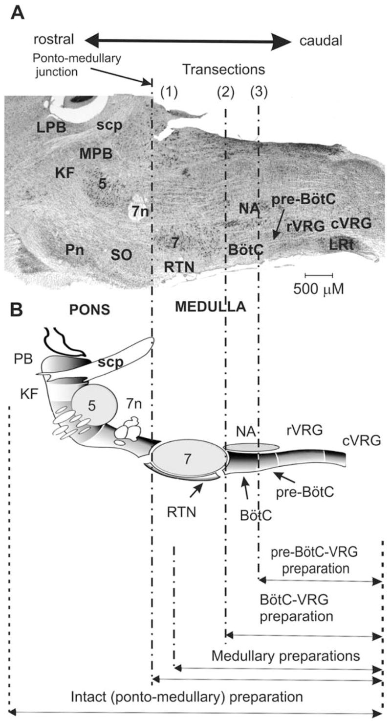
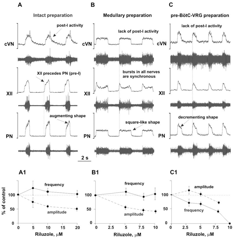
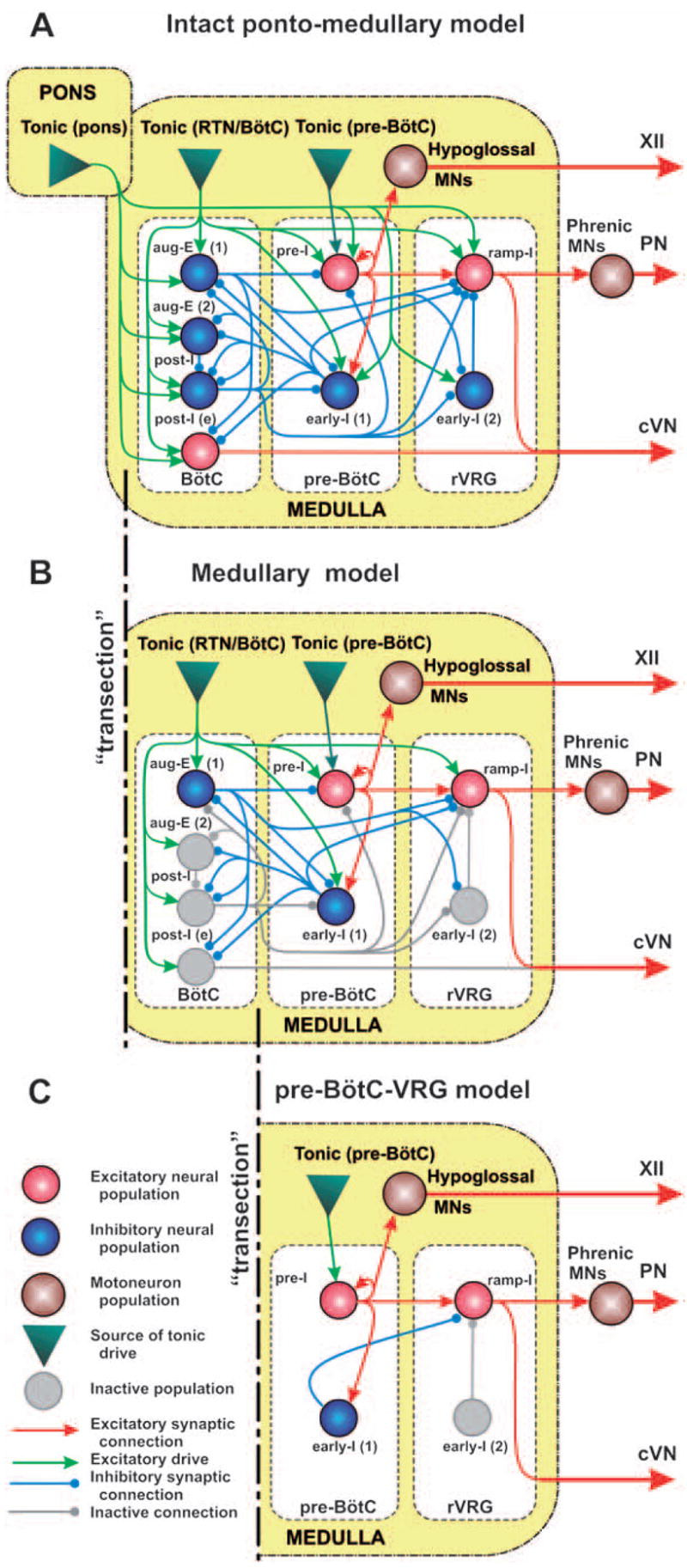
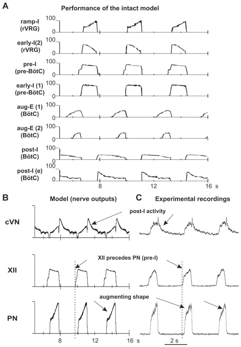
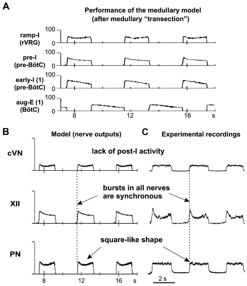
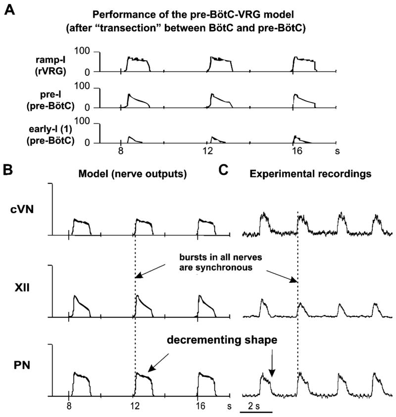
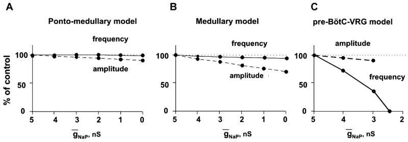
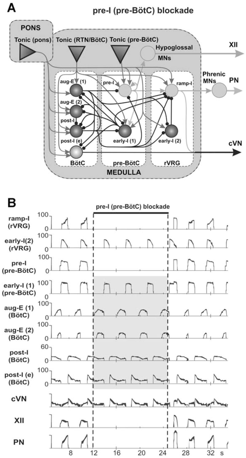
The brainstem respiratory network can operate in multiple functional states engaging different state-dependent neural mechanisms. These mechanisms were studied in the in situ perfused rat brainstem-spinal cord preparation using sequential brainstem transections and administration of riluzole, a pharmacological blocker of persistent sodium current (INaP). Dramatic transformations in the rhythmogenic mechanisms and respiratory motor pattern were observed after removal of the pons and subsequent medullary transactions down to the rostral end of pre-Bötzinger complex (pre-BötC). A computational model of the brainstem respiratory network was developed to reproduce and explain these experimental findings. The model incorporates several interacting neuronal compartments, including the ventral respiratory group (VRG), pre-BötC, Bötzinger complex (BötC), and pons. Simulations mimicking the removal of circuit components following transections closely reproduce the respiratory motor output patterns recorded from the intact and sequentially reduced brainstem preparations. The model suggests that both the operating rhythmogenic mechanism (i.e., network-based or pacemaker-driven) and the respiratory pattern generated (e.g., three-phase, two-phase, or one-phase) depend on the state of the pre-BötC (expression of INaP-dependent intrinsic rhythmogenic mechanisms) and the BötC (providing expiratory inhibition in the network). At the same time, tonic drives from pons and multiple medullary chemoreceptive sites appear to control the state of these compartments and hence the operating rhythmogenic mechanism and motor pattern. Our results suggest that the brainstem respiratory network has a spatial (rostral-to-caudal) organization extending from the rostral pons to the VRG, in which each functional compartment is controlled by more rostral compartments. The model predicts a continuum of respiratory network states relying on different contributions of intrinsic cellular properties versus synaptic interactions for the generation and control of the respiratory rhythm and pattern.


Figures

Fig. 1
Fig. 1
Parasagittal view of rodent brainstem (section through the level of the compact part of nucleus ambiguus) and spatially arrayed compartments of respiratory CPG network. (A) Respiratory-related ponto-medullary regions in the mature rat brainstem with several transection planes (dot-dashed lines) used in experimental studies. (B) Corresponding schematic diagram of respiratory-related brainstem compartments in parasagittal section of rat brain (created by George Alheid and used with permission) with transactions and resultant reduced preparations indicated at the bottom. Abbreviations: 5 – trigeminal nucleus; 7 — facial nucleus; 7n — facial nerve; BötC — Bötzinger Complex; cVRG — caudal ventral respiratory group; KF —Kölliker–Fuse nucleus; LPB — lateral parabrachial nucleus; LRt: lateral reticular nucleus; MPB — medial parabrachial nucleus; NA — nucleus ambiguus; PB — parabrachial nuclei; Pn —pontine nuclei; pre-BötC — pre-Bötzinger Complex; RTN —retrotrapezoid nucleus; rVRG — rostral ventral respiratory group; scp — superior cerebellar peduncle; SO — superior olive.
Fig. 2
Fig. 2
(A–C) Activity patterns of phrenic (PN), hypoglossal (XII), and central vagus (cVN) nerves from the intact (A), medullary (B), and pre-BötC–VGR (C) preparations. Each diagram shows the recorded (bottom trace) and integrated (upper trace) motor output activities. See text for details. (A1–C1) Dose-dependent effects of INaP current blocker riluzole on the frequency (solid lines) and amplitude (dashed lines) of PN bursts in the intact (A1), medullary (B1), and pre-BötC–VGR (C1) preparations. Riluzole in the concentrations shown in horizontal axes was added to the perfusate. Note that the frequency of PN bursts does not significantly change in the intact and medullary preparations (A1 and B1), but dramatically decreases with riluzole concentration in the pre-BötC–VGR preparation and, finally, the PN activity was abolished at riluzole concentration of 10 μM (C1).
Fig. 3
Fig. 3
The schematic of the full (intact) model (A) and the reduced medullary (B) and pre-BötC–VGR (C) models. Neural populations are represented by spheres. Excitatory and inhibitory synaptic connections are shown by arrows and small circles, respectively. Sources of excitatory drives are shown by triangles. All conditional symbols are shown in the left bottom corner. See explanations in the text.
Fig. 4
Fig. 4
Performance of the intact model (network architecture shown in Fig. 3A). (A) Activity of each neural population (labeled on the left) is represented by the histogram of average neuronal spiking frequency (number of spikes per second per neuron, bin = 30 ms). See explanations in the text. (B) Integrated activity of motor (nerve) outputs (PN, XII, and cVN) in this model. (C) Integrated patterns of activity of phrenic (PN), hypoglossal (XII), and central vagus (cVN) nerves obtained from the intact preparation (from Fig. 2A) shown for comparison. See all explanations in the text.
Fig. 5
Fig. 5
Neuronal population activities and motor output patterns of the medullary model (shown in Fig. 3B). (A) Activity (spike frequency histograms) of all neural populations (labeled on the left). See explanations in the text. (B) Integrated activity of motor (nerve) outputs (PN, XII, and cVN) in this model. (C) Integrated patterns of activity of phrenic (PN), hypoglossal (XII), and central vagus (cVN) nerves obtained from a medullary preparation (from Fig. 2B) shown for comparison. See explanations in the text.
Fig. 6
Fig. 6
Performance of the pre-BötC–VGR model (shown in Fig. 3C). (A) Activity of all neural populations (labeled on the left). See explanations in the text. (B) Integrated activity of motor (nerve) outputs (PN, XII, and cVN) in this model. (C) Integrated patterns of activity of phrenic (PN), hypoglossal (XII), and central vagus (cVN) nerves obtained from a pre-BötC–VGR preparation (from Fig. 2C) shown for comparison. See all explanations in the text.
Fig. 7
Fig. 7
Effect of reduction of maximum conductance for the persistent sodium channels (NaP) in all neurons of the pre-I population of pre-BötC on frequency (solid lines) and amplitude (dashed lines) of PN bursts in the intact (A), medullary (B), and pre-BötC–VGR (C) models. Note that the frequency of PN bursts does not change in the intact and medullary models (A and B), but dramatically decreases with the reduction of NaP in the pre-BötC–VGR model and, finally, the PN activity is abolished at NaP = 2.5 nS (C). Compare with the corresponding graphs in Fig. 2A1–C1. See explanations in the text.
Fig. 8
Fig. 8
Neuronal activity patterns in the intact model (Fig. 3A) when the activity of the pre-I population of pre-BötC is suppressed. Activity of pre-I population was suppressed by setting the maximal conductance for fast sodium current to zero for the period shown by a horizontal bar at the top. During this period the pre-I population of pre-BötC as well as the phrenic (PN) and hypoglossal (XII) nerves show no activity. At the same time, an “expiratory” rhythm continues despite of the blockade of inspiration.

References

    1. Abdala APL, Koizumi H, Rybak IA, Smith JC, Paton JFR. The 3-2-1 state respiratory rhythm generator hypothesis revealed by microsectioning, reduced extracellular chloride and alterations in arterial gas tensions in the in situ rat. Exp Biology Abstr. 2007:610.4.
    1. Alheid GF, Milsom WK, McCrimmon DR. Pontine influences on breathing: an overview. Respir Physiol Neurobiol. 2004;143:105–114. - PubMed
    1. Butera RJ, Rinzel JR, Smith JC. Models of respiratory rhythm generation in the pre-Bötzinger complex: I. Bursting pacemaker neurons. J Neurophysiol. 1999a;82:382–397. - PubMed
    1. Butera RJ, Rinzel JR, Smith JC. Models of respiratory rhythm generation in the pre-Bötzinger complex: II. Populations of coupled pacemaker neurons. J Neurophysiol. 1999b;82:398–415. - PubMed
    1. Cohen MI. Neurogenesis of respiratory rhythm in the mammal. Physiol Rev. 1979;59:1105–1173. - PubMed

Publication types

LinkOut - more resources