. 2007 Dec;13(12):2141-50.
doi: 10.1261/rna.678307. Epub 2007 Oct 9.

In vitro dimerization of human immunodeficiency virus type 1 (HIV-1) spliced RNAs

Affiliations

In vitro dimerization of human immunodeficiency virus type 1 (HIV-1) spliced RNAs

Lucile Sinck et al. RNA. 2007 Dec.

Abstract

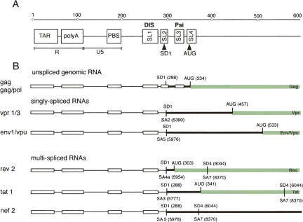
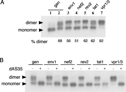
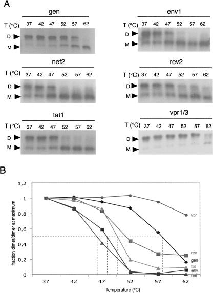
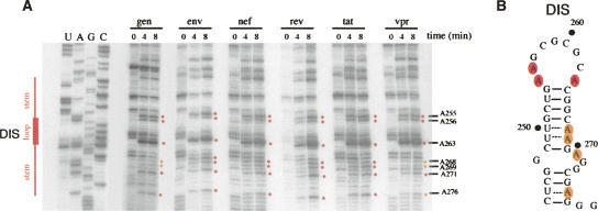
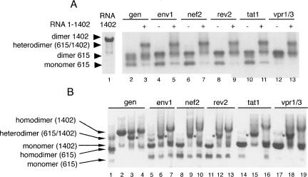
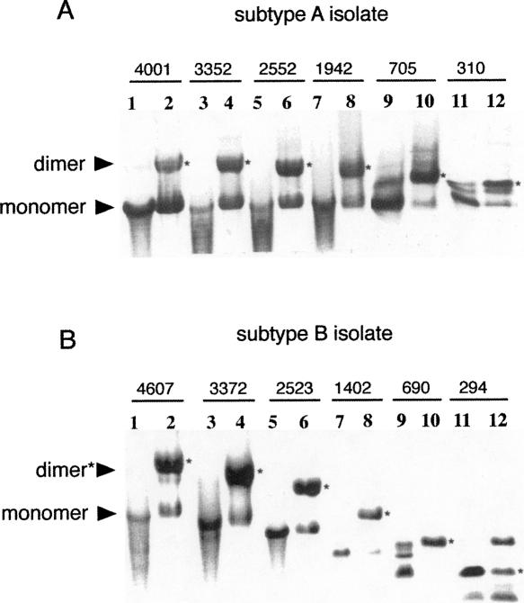
The human immunodeficiency virus type 1 (HIV-1) packages its genomic RNA as a dimer of homologous RNA molecules that has to be selected among a multitude of cellular and viral RNAs. Interestingly, spliced viral mRNAs are packaged into viral particles with a relatively low efficiency despite the fact that they contain most of the extended packaging signal found in the 5' untranslated region of the genomic RNA, including the dimerization initiation site (DIS). As a consequence, HIV-1 spliced viral RNAs can theoretically homodimerize and heterodimerize with the genomic RNA, and thus they should directly compete with genomic RNA for packaging. To shed light on this issue, we investigated for the first time the in vitro dimerization properties of spliced HIV-1 RNAs. We found that singly spliced (env, vpr) and multispliced (tat, rev, and nef) RNA fragments are able to dimerize in vitro, and to efficiently form heterodimers with genomic RNA. Chemical probing experiments and inhibition of RNA dimerization by an antisense oligonucleotide directed against the DIS indicated that the DIS is structurally functional in spliced HIV-1 RNA, and that RNA dimerization occurs through a loop-loop interaction. In addition, by combining in vitro transcription and dimerization assays, we show that heterodimers can be efficiently formed only when the two RNA fragments are synthesized simultaneously, in the same environment. Together, our results support a model in which RNA dimerization would occur during transcription in the nucleus and could thus play a major role in splicing, transport, and localization of HIV-1 RNA.


Figures

FIGURE 1.
FIGURE 1.
Genomic and spliced HIV-1 (NL4.3) RNAs. (A) Scheme of the first 600 nt of HIV-1 genomic RNA. (R) repeat sequence, (TAR) trans-acting responsive element, (polyA) 5′ copy of the polyadenylation signal, (U5) unique sequence at the 5′ end of the RNA genome, (PBS) primer-binding site, (DIS) dimerization initiation site, (SD) major splice-donor site, (PSI) major packaging signal, and (AUG) translation initiator codon of gag gene. (B) Spliced and genomic RNAs used in this study. (Open boxes) The different RNA elements, (black thick lines) untranslated regions, and (green thick lines) coding regions. The positions of splice donor (SD) and acceptor (SA) sites are indicated above and under the genomic RNA, respectively.
FIGURE 2.
FIGURE 2.
In vitro dimerization of spliced HIV-1 RNAs. (A) RNA fragments were incubated in dimer buffer (high-salt conditions) and run on a native 0.8% agarose gel (TB 0.5×, 0.1 mM MgCl2). Control monomeric RNA (low-salt conditions) is shown only for the genomic RNA fragment (lane 1). Monomers and dimers are indicated, as well as the percentage of dimer formed for each RNA. (B) Inhibition of RNA dimerization. RNAs were incubated in high-salt conditions in the absence (−) or presence (+) of an antisense oligodeoxynucleotide (dAS35) complementary to the DIS stem–loop.
FIGURE 3.
FIGURE 3.
Thermal stability of genomic and spliced 1–615 RNA dimers. (A) After RNA dimerization in high-salt buffer at 37°C, samples were incubated at different temperatures ranging from 37°C to 62°C and loaded on 0.8% native agarose gels. Gels were fixed, dried, and autoradiographed. (B) Thermal stability curves. Dimer yields were normalized relative to the dimer yield at 37°C and plotted as a function of temperature.
FIGURE 4.
FIGURE 4.
Dimethylsulfate (DMS) probing of the Dimerization Initiation Site (DIS) region of spliced and genomic HIV-1 RNAs. (A) RNAs were modified with DMS, and positions of methylated bases were revealed by primer extension before loading on an 8% polyacrylamide denaturing gel. Lanes U,A,C,G correspond to the sequence of gRNA. DIS and SD positions are indicated. Red dots represent the three purines flanking the self-complementary sequence of the DIS self-complementary sequence. (B) Secondary structure of the DIS. Nucleotides modified by DMS are indicated in red (highly reactive) or in orange (moderately reactive).
FIGURE 5.
FIGURE 5.
Heterodimerization of genomic and spliced HIV-1 RNAs. (A) Heterodimerization with purified RNA fragments. Radiolabeled 1–615 RNAs were incubated in high-salt buffer in the absence (−) or in presence (+) of genomic RNA fragment 1–1402. (Lane 1) The 1–1402 RNA dimer visualized by UV. Positions of monomer, homodimer, and heterodimer species are indicated. (B) Heterodimer formation during in vitro transcription. DNA templates generating 1–615 spliced or genomic RNA fragments were submitted to transcription either alone (lanes 1,2,5,8,11,14,17) or with template generating genomic RNA fragment 1–1402 (lanes 3,6,9,12,15,18). RNAs 1–615 and 1–1402 were also transcribed separately before being mixed together for another 30-min incubation at 37°C (lanes 4,7,10,13,16,19). Samples were analyzed on a native ethidium-bromide-containing agarose gel. (Gray star) The 1–615/1–1402 heterodimer, (lane 1) genomic RNA 1–615, (lane 2) genomic RNA 1–1402. The names of each RNA are indicated at the top of the gel. (gen) gRNA.
FIGURE 6.
FIGURE 6.
In vitro dimerization of long subtype A (A) and subtype B (B) RNA fragments during transcription. Samples were analyzed on native agarose ethidium-bromide-containing gels directly after in vitro run-off transcription. (Black stars) Dimers. The size of each RNA fragment is indicated at the top of the gels. (Odd lanes) RNA fragments that have been denatured 2 min at 80°C before loading, (even lanes) not heat-treated. RNA fragments are staggered in panel B (compared with panel A) due to direct loading of samples after transcription. In this case, RNAs migrated according to their length. In panel A, RNA samples have been loaded at different time points to place the RNA bands at the same position in the gel.

Similar articles

Cited by

References

    1. Aldovini, A., Young, R.A. Mutations of RNA and protein sequences involved in human immunodeficiency virus type 1 packaging result in production of noninfectious virus. J. Virol. 1990;64:1920–1926. - PMC - PubMed
    1. Andersen, E.S., Jeeninga, R.E., Damgaard, C.K., Berkhout, B., Kjems, J. Dimerization and template switching in the 5′ untranslated region between various subtypes of human immunodeficiency virus type 1. J. Virol. 2003;77:3020–3030. - PMC - PubMed
    1. Balakrishnan, M., Fay, P.J., Bambara, R.A. The kissing hairpin sequence promotes recombination within the HIV-I 5′ leader region. J. Biol. Chem. 2001;276:36482–36492. - PubMed
    1. Balakrishnan, M., Roques, B.P., Fay, P.J., Bambara, R.A. Template dimerization promotes an acceptor invasion-induced transfer mechanism during human immunodeficiency virus type 1 minus-strand synthesis. J. Virol. 2003;77:4710–4721. - PMC - PubMed
    1. Basyuk, E., Boulon, S., Skou Pedersen, F., Bertrand, E., Vestergaard Rasmussen, S. The packaging signal of MLV is an integrated module that mediates intracellular transport of genomic RNAs. J. Mol. Biol. 2005;354:330–339. - PubMed

Publication types