

Review
doi: 10.3174/ajnr.A0781.
Epub 2007 Oct 9.
Neuropathology for the neuroradiologist: plaques and tangles
Affiliations
- PMID: 17925367
- PMCID: PMC8119079
- DOI: 10.3174/ajnr.A0781
Item in Clipboard
Review
Neuropathology for the neuroradiologist: plaques and tangles
AJNR Am J Neuroradiol.
2008 Jan.
Abstract
Histologically identified intracellular and extracellular inclusions and structures often provide a tissue diagnosis of a specific disease process. Moreover, these deposits may provide clues about the pathogenesis of the disease in which they are found. Two distinctive structures seen within the brains of patients clinically diagnosed with dementia of the Alzheimer type are extracellular plaques and intracellular neurofibrillary tangles. The purpose of this report is to review the significance of plaques and neurofibrillary tangles in the context of Alzheimer disease.
Figures
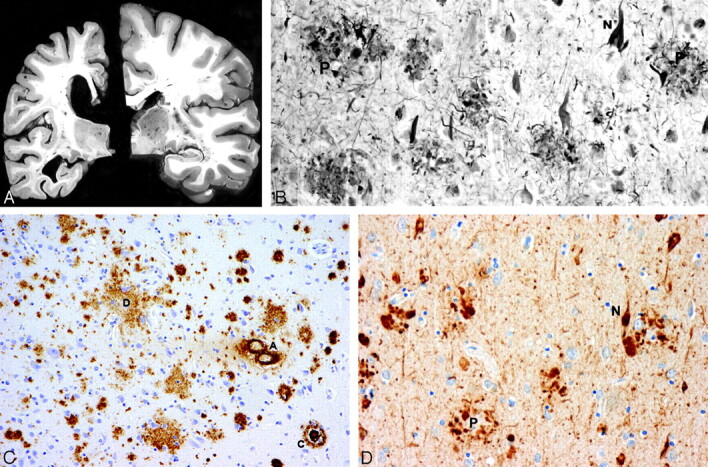
A, Atrophy of the brain. On the left, a section of the hemibrain of a 70-year-old patient with AD and, on the right, a healthy aged control brain. The AD brain shows marked atrophy, dilation of the lateral ventricle, and a small hippocampus. B, Neurofibrillary tangles (N) and neuritic plaques (P) in the hippocampus. Modified Bielschowsky silver impregnation. C, β-amyloidosis in the frontal lobe: a diffuse plaque (D), a cored plaque (C), and cerebral amyloid angiopathy (A). β-amyloid (10D5) immunohistochemistry. D, Neurofibrillary tangles (N) and neuritic plaques (P) in the frontal lobe. Phosphorylated ô immunohistochemistry.
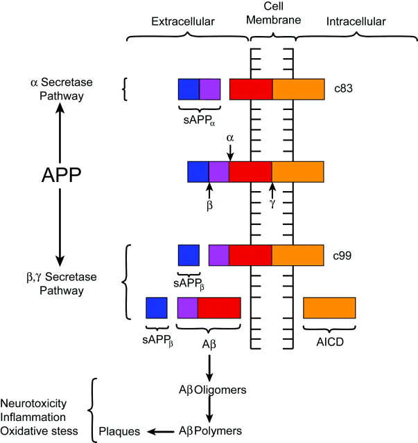
Diagrammatic representation of APP processing. APP is a transmembrane protein with both extra- and intracellular components. APP is processed by 2 competing pathways. The α-secretase pathway generates sAPPα and C83 protein by cleavage of the α-secretase enzyme (α). In the β-secretase pathway, the enzyme β-secretase (β) cleaves APP into an sAPPβ fragment and a C99 fragment. The C99 fragment is further cleaved by γ-secretase enzyme (γ) into an amyloid β fragment (Aβ) and an AICD fragment. The Aβ fragments polymerize. The oligomers and polymers exhibit neurotoxicity. As polymerization proceeds to more complex forms, senile plaques are developed. The C83 fragment is also further processed. However, the function of its products is not fully understood. (The relative sizes of the protein fragments are not drawn to scale.)
References
-
- Möller HJ, Graeber MB. The case described by Alois Alzheimer in 1911: historical and conceptual perspectives based on the clinical record and neurohistological sections. Eur Arch Psychiatry Clin Neurosci 1998;248:111–22 - PubMed
-
- Kircher T, Wormstall H. Alois Alzheimer (1864-1915): student days and first scientific activities. J Geriatr Psychiatry Neurol 1997;10:127–29 - PubMed
-
- Brannon WL. Alois Alzheimer (1864–1915). I. Contributions to neurology and psychiatry. J S C Med Assoc 1994;90:399–401 - PubMed
-
- Beach TG. The history of Alzheimer's disease: three debates. J Hist Med Allied Sci 1987;42:327–49 - PubMed
-
- Small DH, Cappai R. Alois Alzheimer and Alzheimer's disease: a centennial perspective. J Neurochem 2006;99:708–10 - PubMed
Publication types
MeSH terms
Grants and funding
LinkOut - more resources
Full Text Sources
Medical