. 2007 Oct 16;104(42):16410-5.
doi: 10.1073/pnas.0705759104. Epub 2007 Oct 9.

Hierarchies, multiple energy barriers, and robustness govern the fracture mechanics of alpha-helical and beta-sheet protein domains

Affiliations

Hierarchies, multiple energy barriers, and robustness govern the fracture mechanics of alpha-helical and beta-sheet protein domains

Theodor Ackbarow et al. Proc Natl Acad Sci U S A. .

Abstract

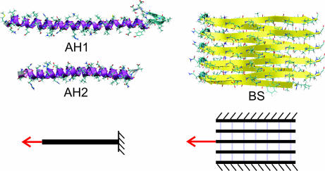
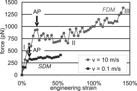
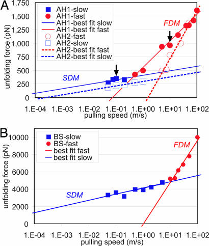
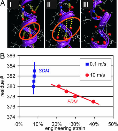
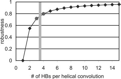
The fundamental fracture mechanisms of biological protein materials remain largely unknown, in part, because of a lack of understanding of how individual protein building blocks respond to mechanical load. For instance, it remains controversial whether the free energy landscape of the unfolding behavior of proteins consists of multiple, discrete transition states or the location of the transition state changes continuously with the pulling velocity. This lack in understanding has thus far prevented us from developing predictive strength models of protein materials. Here, we report direct atomistic simulation that over four orders of magnitude in time scales of the unfolding behavior of alpha-helical (AH) and beta-sheet (BS) domains, the key building blocks of hair, hoof, and wool as well as spider silk, amyloids, and titin. We find that two discrete transition states corresponding to two fracture mechanisms exist. Whereas the unfolding mechanism at fast pulling rates is sequential rupture of individual hydrogen bonds (HBs), unfolding at slow pulling rates proceeds by simultaneous rupture of several HBs. We derive the hierarchical Bell model, a theory that explicitly considers the hierarchical architecture of proteins, providing a rigorous structure-property relationship. We exemplify our model in a study of AHs, and show that 3-4 parallel HBs per turn are favorable in light of the protein's mechanical and thermodynamical stability, in agreement with experimental findings that AHs feature 3.6 HBs per turn. Our results provide evidence that the molecular structure of AHs maximizes its robustness at minimal use of building materials.


Conflict of interest statement

The authors declare no conflict of interest.

Figures

Fig. 1.
Fig. 1.
Atomistic geometries of the three protein domains studied here (AH1, AH2, and BS). Surrounding water molecules are not shown for reasons of clarity. The lower part of the plot indicates the boundary conditions (tensile loading for AH1 and AH2 and shear loading for BS). The BS structure consists of two stacks of β-sheets in the out-of-plane direction.
Fig. 2.
Fig. 2.
Examples for force–extension curves of AH1. The fast deformation mode (FDM) is represented by a curve taken at a pulling speed of 10 m/s. The slow deformation mode (SDM) is represented by a pulling experiment at 0.1 m/s. The force–extension behavior consists of three regimes: (I) linear increase in strain until the AP is reached (indicated by arrows) when the first HBs rupture, leading to unfolding of one helical turn; (II) plateau of approximately constant force, during which unfolding of the entire protein occurs; and (III) strain hardening (only partly shown for the FDM).
Fig. 3.
Fig. 3.
Unfolding force of single AHs from the vimentin coiled-coil dimers (A) and a BS amyloid domain (B), as a function of varying pulling speed over four orders of magnitude, ranging from 0.05 to 100 m/s. The results clearly reveal a change in protein-unfolding mechanism from the FDM to the SDM. The arrows in A indicate the representative pulling speeds used for the analysis reported in Figs. 2 and 4.
Fig. 4.
Fig. 4.
Atomistic details of the unfolding mechanism of AH1 in the SDM (AP in Fig. 1 for v = 0.1 m/s). (A) Atomistic representation of the rupture dynamics. The time interval between these snapshots is 20 ps (between I and II) and 40 ps (between II and III). After 20 ps (I to II), all three HBs have ruptured simultaneously, leading to local unfolding of the protein in the next 40 ps (II to III). These snapshots strongly support the concept of cooperative bond rupture in the SDM. Surrounding water molecules are not shown for reasons of clarity. (B) Rupture sequence of the first four HBs as a function of the applied strain [residue number represents the amino acid of the O atom (H acceptor)]. In the FDM, HBs rupture one by one, whereas in the SDM, several HBs rupture almost simultaneously, within 20 ps. In the FDM, the unfolding wave runs from the pulled residue in the direction of the fixed residue, whereas in the SDM, the unfolding “wave” runs in the opposite direction, nucleating at a random residue within the protein sequence.
Fig. 5.
Fig. 5.
Robustness of an AH as a function of parallel HBs per turn, b, predicted by the hierarchical Bell model. Robustness is defined as the ratio of strength of a failed system and an intact system. In the intact system, all HBs contribute to strength, whereas in the failed system, all except one HB contribute to the strength. The shaded bar indicates the number of parallel HBs per turn (3.6 HBs) as observed in nature. This particular molecular geometry corresponds to a robustness value of ≈80%, indicating that the AH is efficient in Pareto's sense (38, 39).

References

    1. Alberts B, Johnson A, Lewis J, Raff M, Roberts K, Walter P. Molecular Biology of the Cell. 4th Ed. New York: Garland; 2002.
    1. Gruber M, Lupas AN. Trends Biochem Sci. 2003;28:679–685. - PubMed
    1. Moir RD, Spann TP. Cell Mol Life Sci. 2001;58:1748–1757. - PMC - PubMed
    1. Wilson KL, Zastrow MS, Lee KK. Cell. 2001;104:647–650. - PubMed
    1. Luhrs T, Ritter C, Adrian M, Riek-Loher D, Bohrmann B, Doeli H, Schubert D, Riek R. Proc Natl Acad Sci USA. 2005;102:17342–17347. - PMC - PubMed

Publication types

LinkOut - more resources