. 2007 Oct 10;2(10):e1004.
doi: 10.1371/journal.pone.0001004.

Tsukushi modulates Xnr2, FGF and BMP signaling: regulation of Xenopus germ layer formation

Affiliations

Tsukushi modulates Xnr2, FGF and BMP signaling: regulation of Xenopus germ layer formation

Samantha A Morris et al. PLoS One. .

Abstract

Background: Cell-cell communication is essential in tissue patterning. In early amphibian development, mesoderm is formed in the blastula-stage embryo through inductive interactions in which vegetal cells act on overlying equatorial cells. Members of the TGF-beta family such as activin B, Vg1, derrière and Xenopus nodal-related proteins (Xnrs) are candidate mesoderm inducing factors, with further activity to induce endoderm of the vegetal region. TGF-beta-like ligands, including BMP, are also responsible for patterning of germ layers. In addition, FGF signaling is essential for mesoderm formation whereas FGF signal inhibition has been implicated in endoderm induction. Clearly, several signaling pathways are coordinated to produce an appropriate developmental output; although intracellular crosstalk is known to integrate multiple pathways, relatively little is known about extracellular coordination.

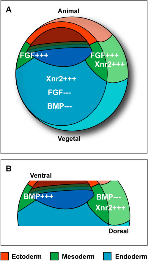
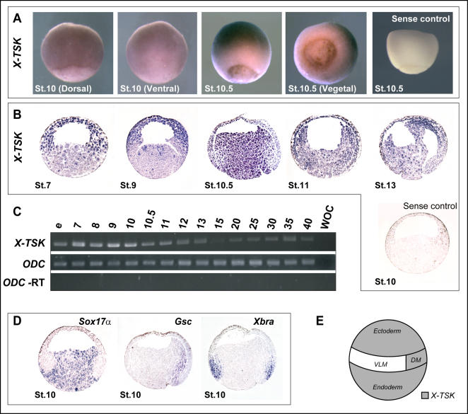
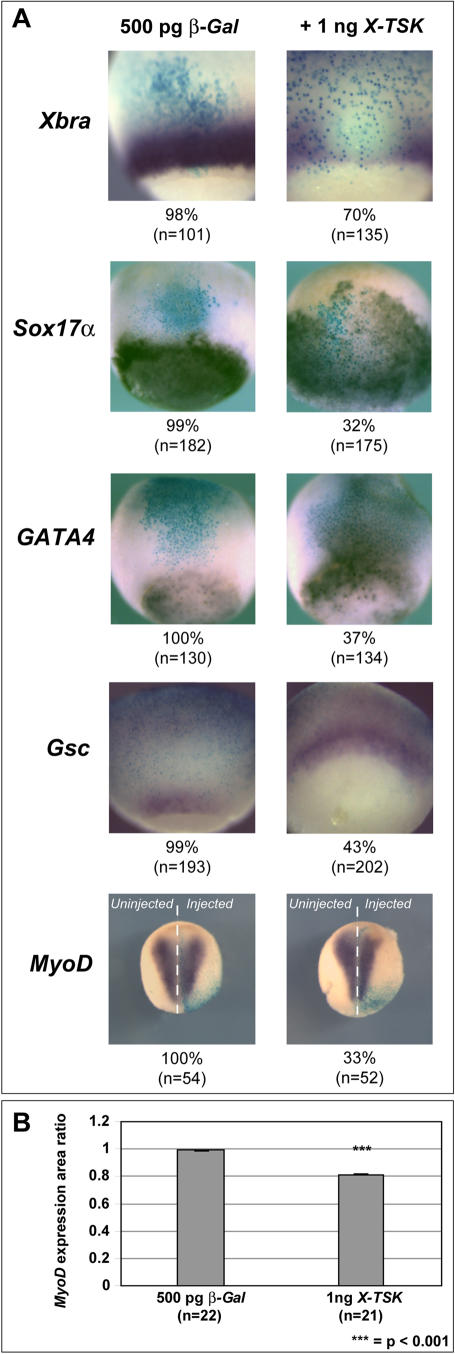
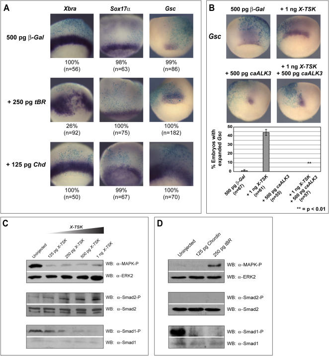
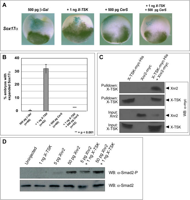
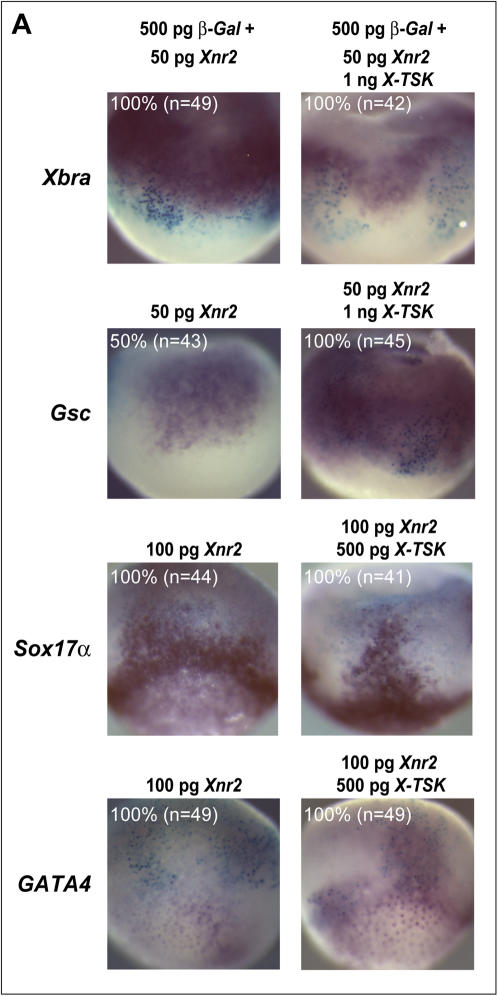
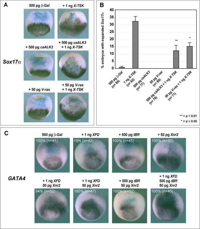
Methodology/principal findings: Here, we show that Xenopus Tsukushi (X-TSK), a member of the secreted small leucine rich repeat proteoglycan (SLRP) family, is expressed in ectoderm, endoderm, and the organizer during early development. We have previously reported that X-TSK binds to and inhibits BMP signaling in cooperation with chordin. We now demonstrate two novel interactions: X-TSK binds to and inhibits signaling by FGF8b, in addition to binding to and enhancement of Xnr2 signaling. This signal integration by X-TSK at the extracellular level has an important role in germ layer formation and patterning. Vegetally localized X-TSK potentiates endoderm formation through coordination of BMP, FGF and Xnr2 signaling. In contrast, X-TSK inhibition of FGF-MAPK signaling blocks ventrolateral mesoderm formation, while BMP inhibition enhances organizer formation. These actions of X-TSK are reliant upon its expression in endoderm and dorsal mesoderm, with relative exclusion from ventrolateral mesoderm, in a pattern shaped by FGF signals.

Conclusions/significance: Based on our observations, we propose a novel mechanism by which X-TSK refines the field of positional information by integration of multiple pathways in the extracellular space.


Conflict of interest statement

Competing Interests: The authors have declared that no competing interests exist.

Figures

Figure 1
Figure 1. Signaling involved in Xenopus germ layer formation.
(A) Selected signaling pathways involved in Xenopus mesoderm and endoderm formation. Activation of pathways indicated by ‘+++’, inhibition of pathways indicated by ‘−−−‘. Ectoderm = red, mesoderm = green, endoderm = blue. FGF signal activity is required for mesoderm formation in addition to activity of activin-like signaling (represented here by Xnr2). FGF and BMP signal inhibition with Xnr2 signal activation is involved in endoderm induction mechanisms. (B) Selected signaling pathways involved in Xenopus mesoderm patterning. Active BMP signaling produces mesoderm with ventral character, whereas inhibition of BMP signaling produces mesoderm of dorsal character. Also, Xnr2 expressed in the dorsal region has activity to induce dorsal mesoderm.
Figure 2
Figure 2. X-TSK expression in Xenopus.
(A) Whole mount in situ hybridization of X-TSK in Xenopus gastrula stage embryos, including sense control. Purple staining indicates X-TSK expression. Orientations and stages as indicated. X-TSK is expressed in dorsal marginal zone (DMZ) and ectoderm from stage 10, and endoderm from stage 10.5. (B) In situ hybridization of X-TSK in sectioned Xenopus embryos, including sense control. Orientation: animal top, vegetal bottom, dorsal right, stages as indicated. X-TSK is expressed maternally (stage 7) in the animal region, with light staining in the vegetal region. From stage 10.5, X-TSK expression is detected throughout the endoderm. (C) Expression levels of X-TSK (upper panel) measured by RT-PCR from egg to stage 41, including ODC expression (middle panel) and -RT control (lower panel). WOC = Water Only Control. X-TSK is expressed at highest levels during germ layer formation and gastrulation. (D) Comparative expression of Sox17α (marking endoderm), Gsc (dorsal mesoderm), and Xbra (pan-mesoderm) in sectioned stage 10 embryos. (E) Schematic of X-TSK expression (grey) in ectoderm, dorsal mesoderm and endoderm.
Figure 3
Figure 3. Loss of X-TSK function.
(A) In situ hybridization of endoderm markers, Sox17α (upper row), and GATA4 (lower row) in sectioned early gastrula (stage 10) embryos, purple staining indicates expression. Orientation: animal top, vegetal bottom. All embryos injected with 500 pg β-Galactosidase (β-Gal) to identify targeted area (blue staining), with 20 ng control morpholino (CMO) or 20 ng X-TSK morpholino (XMO). Endoderm marker staining is reduced in XMO injected embryos, as indicated by general loss of purple staining (Sox17α) and loss of punctate staining (GATA4), detailed in the zoomed panel. Rescues were performed with 1 ng H-TSK, or 50 pg Xnr2, restoring endoderm marker expression. Detailed analysis of GATA4 staining in sectioned embryos. Numbers of GATA4 foci were counted, as represented graphically, relative to uninjected control. XMO injection reduces GATA4 foci by 50% (p = <0.001), partially rescued by 1 ng H-TSK and 50 pg Xnr2 to over 80% relative to control (p = <0.001). (B) Whole mount in situ hybridization of dorsal mesoderm marker, Gsc in stage 10.5 embryos (dorsal orientation) and MyoD in stage 16 (anterior top, posterior bottom) in embryos injected with 500 pg β-Gal, with 20 ng CMO or 20 ng XMO. Gsc expression is reduced in XMO injected embryos, whereas MyoD expression is expanded by 30% (relative to control, p = <0.001) on the injected side, as identified by blue β-Gal staining. (C) Gut morphology in stage 40 embryos injected with 20 ng CMO or 20 ng XMO. Gut width is reduced by 21% in XMO injected embryos, relative to uninjected embryos (p = <0.001).
Figure 4
Figure 4. Gain of X-TSK function.
(A) Whole mount in situ hybridization of germ layer markers in embryos injected with 500 pg β-Gal and 1 ng X-TSK, with percentage occurrence of demonstrated phenotype and ‘n’ numbers indicated below images. Xbra (pan-mesoderm) expression is inhibited and Sox17α and GATA4 (endoderm) expression is expanded, stage 10.5, lateral orientation. Gsc (dorsal mesoderm) expression is expanded, stage 10.5, dorsal orientation. MyoD expression is inhibited on the injected side, as identified by blue β-Gal staining, stage 16, anterior top, posterior bottom. (B) Graphical representation of MyoD expression in (A). MyoD expression is reduced by 20% on the injected side (p = <0.001).
Figure 5
Figure 5. Mechanism of X-TSK function: signal analysis.
(A) Whole mount in situ hybridization of germ layer markers in embryos injected with 500 pg β-Gal with 250 pg truncated BMP receptor (tBR) or 125 pg Chordin (Chd), with percentage occurance of demonstrated phenotype and ‘n’ numbers. Xbra and Sox17α phenotypes differ in comparison to X-TSK overexpression, whereas Gsc expression is commonly expanded. (B) Whole mount in situ hybridization of Gsc in embryos injected with 500 pg β-Gal with 1 ng X-TSK, 500 pg caALK3 and X-TSK with caALK3, dorsal orientation. caALK3 blocks X-TSK mediated expansion of Gsc expression. (C) Western blotting of MAPK and Smad1 phosphorylation in animal caps and Smad2 phosphorylation in DMZ explants, with total MAPK, Smad2 and Smad1 controls in explants injected with X-TSK (125 pg-1 ng) (D) 125 pg Chd or 250 pg tBR. X-TSK inhibits MAPK and BMP phosphorylation in animal caps whilst activating Smad2 phosphorylation in DMZ. Chd and tBR similarly inhibit BMP phosphorylation, but contrast with X-TSK in MAPK and Smad2 phosphorylation status.
Figure 6
Figure 6. X-TSK inhibition and binding of FGF8b.
(A) Western blotting of MAPK phosphorylation in animal caps injected with 20–40 ng CMO and 20–40 ng XMO. Depletion of X-TSK with XMO activates MAPK phosphorylation. (B) Semi-quantitative RT-PCR of Xbra expression in DMZ injected with 20 ng CMO, 20 ng XMO, 500 pg XFD and 500 pg XFD with 20 ng XMO. WE = Whole embryo, WOC = Water only control. Inhibition of FGF signals with XFD blocks Xbra expression activated upon depletion of X-TSK with XMO. (C) Whole mount in situ hybridization of Xbra in stage 10.5 embryos, lateral orientation. Embryos injected with 500 pg β-Gal with 1 ng X-TSK, 50 pg V-ras and X-TSK with V-ras. V-ras blocks X-TSK mediated inhibition of Xbra expression in 100% of embryos analyzed (p = <0.01), represented graphically in (D). (E) Western blotting of MAPK phosphorylation in animal caps injected with X-TSK and V-ras. V-ras blocks X-TSK mediated inhibition of MAPK phosphorylation. (F) Western blotting of MAPK phosphorylation in animal caps injected with X-TSK and iFGFR, in the presence or absence of chemical dimerisation agent, AP20187. Induced dimerisation blocks the activity of X-TSK to inhibit MAPK phosphorylation. (G) Western blotting of MAPK phosphorylation in animal caps injected with X-TSK and FGF8b. X-TSK inhibits MAPK phosphorylation activated by FGF8b. (H) Western blotting of nickel bead pulldown of FGF8b-FLAG in complex with X-TSK-Myc-His. Top panel: detection of FGF8b-FLAG in complex with X-TSK-Myc-His (third lane). Second panel: detection of X-TSK-Myc-His pulled down. Third and bottom panels: detection of FGF8b-FLAG and X-TSK-Myc-His input into the pulldown reaction.
Figure 7
Figure 7. X-TSK requires intact Xnr signaling for endoderm induction; X-TSK binds to and enhances Xnr2 Signaling.
(A) Whole mount in situ hybridization of Sox17α in embryos injected with 500 pg β-Gal with 1 ng X-TSK, 500 pg CerS and 500 pg CerS with 1 ng X-TSK, lateral orientation. (B) Introduction of CerS blocks X-TSK expansion of Sox17α in 100% of embryos analyzed (p = <0.001). (C) Western blotting of nickel bead pulldown of Xnr2-Myc in complex with X-TSK-Myc-His. Top panel: detection of Xnr2-Myc in complex with X-TSK-Myc-His (third lane). Second panel: detection of X-TSK-Myc-His pulled down. Third and bottom panels: detection of Xnr2-Myc and X-TSK-Myc-His input into the pulldown reaction. (D) Western blotting of Smad2 phosphorylation in animal caps injected with 1 ng X-TSK, 5 pg and 50 pg Xnr2. X-TSK enhances Smad2 phosphorylation, particularly evident with 5 pg Xnr2.
Figure 8
Figure 8. X-TSK changes local response to Xnr2.
(A) Whole mount in situ hybridization of Xbra, Gsc (dorsal orientation) Sox17α and GATA4 in embryos injected with 500 pg β-Gal with 50 pg Xnr2, and 50 pg Xnr2 with 1ng X-TSK and 500 pg X-TSK, lateral orientation. Xbra expression is not detected in Xnr2-X-TSK expressing cells, as identified by β-Gal staining. Xnr2 mediated expansion of Gsc expression is enhanced by X-TSK. Expression of endoderm markers Sox17α and GATA4 expanded by Xnr2 is enhanced by X-TSK, suggesting that X-TSK changes local cellular response to Xnr2.
Figure 9
Figure 9. BMP and FGF signal activation blocks X-TSK mediated endoderm induction: triple signal regulation.
(A) Whole mount in situ hybridization of Sox17α in embryos injected with 500 pg β-Gal with 1 ng X-TSK, 500 pg caALK3, 50 pg V-ras and 500 pg caALK3, 50 pg V-ras with 1 ng X-TSK, lateral orientation. (B) Graphic representation of quantity of embryos demonstrating expanded Sox17α expression. Introduction of caALK3 and V-ras partially blocks X-TSK expansion of Sox17α (p = 0.01 and 0.05 respectively). (C) Whole mount in situ hybridization of GATA4 in embryos injected with 500 pg β-Gal with combinations of 1 ng XFD, 500 pg tBR, and 50 pg Xnr2, lateral orientation. Demonstrated phenotype frequencies with n-numbers in white text. A triple combination of 1 ng XFD, 500 pg tBR, and 50 pg Xnr2 produces the strongest expansion of GATA4 expression.
Figure 10
Figure 10. Transcriptional regulation of X-TSK and model of X-TSK function in germ layer formation and patterning.
(A) Semi-quantitative RT-PCR of X-TSK expression in animal caps injected with 1 ng XFD, 50 pg V-ras or 50 pg caFGFR. WE = Whole embryo, WOC = Water only control. Inhibition of FGF signals with XFD enhances TSK expression, whereas activation of FGF signals with V-ras or caFGFR reduces TSK expression levels. (B) Model of TSK function in Xenopus germ layer formation and patterning: dorsal-ventral mesoderm patterning. X-TSK in dorsal mesoderm (red) inhibits BMP signaling to promote dorsal mesoderm formation, as marked by Gsc expression. This is possibly also enhanced through activation of Xnr2 signals by TSK. MAPK activation inhibits X-TSK expression in ventrolateral mesoderm, where X-TSK inhibits expression of ventrolateral mesoderm markers such as Xbra, through inhibition of FGF signaling. This network of signaling may contribute to clear patterning of the mesoderm. (C) Model of TSK function in endoderm formation. X-TSK coordinates inhibition of FGF and BMP signals with activation of Xnr2 signaling to induce endoderm formation (green), as marked by Sox17α. Again, X-TSK inhibits expression of ventrolateral mesoderm (blue) markers such as Xbra, through inhibition of FGF signaling and may contribute to the distinction between endoderm and mesoderm specific gene expression.

Similar articles

Cited by

References

    1. Heasman J. Patterning the Xenopus blastula. Development. 1997;124:4179–4191. - PubMed
    1. Darras S, Marikawa Y, Elinson RP, Lemaire P. Animal and vegetal pole cells of early Xenopus embryos respond differently to maternal dorsal determinants: implications for the patterning of the organiser. Development. 1997;124:4275–4286. - PubMed
    1. Loose M, Patient R. A genetic regulatory network for Xenopus mesendoderm formation. Dev Biol. 2004;271:467–478. - PubMed
    1. Smith JC, Price BM, Nimmen KV, Huylebroeck D. Identification of a potent Xenopus mesoderm-inducing factor as a homologue of activin A. Nature. 1990;345:729–731. - PubMed
    1. Weeks DL, Melton DA. A maternal mRNA localized to the vegetal hemisphere in Xenopus eggs codes for a growth factor related to TGF-beta. Cell. 1987;51:861–867. - PubMed

Publication types