

The chemokine receptor CXCR4 strongly promotes neuroblastoma primary tumour and metastatic growth, but not invasion
- PMID: 17925864
- PMCID: PMC1995764
- DOI: 10.1371/journal.pone.0001016
The chemokine receptor CXCR4 strongly promotes neuroblastoma primary tumour and metastatic growth, but not invasion
Abstract
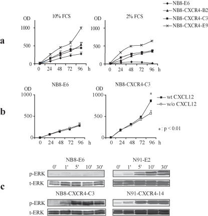
Neuroblastoma (NB) is a heterogeneous, and particularly malignant childhood neoplasm in its higher stages, with a propensity to form metastasis in selected organs, in particular liver and bone marrow, and for which there is still no efficient treatment available beyond surgery. Recent evidence indicates that the CXCR4/CXCL12 chemokine/receptor axis may be involved in promoting NB invasion and metastasis. In this study, we explored the potential role of CXCR4 in the malignant behaviour of NB, using a combination of in vitro functional analyses and in vivo growth and metastasis assessment in an orthotopic NB mouse model. We show here that CXCR4 overexpression in non-metastatic CXCR4-negative NB cells IGR-NB8 and in moderately metastatic, CXCR4 expressing NB cells IGR-N91, strongly increased tumour growth of primary tumours and liver metastases, without altering the frequency or the pattern of metastasis. Moreover shRNA-mediated knock-down experiments confirmed our observations by showing that silencing CXCR4 in NB cells impairs in vitro and almost abrogates in vivo growth. High levels of CXCL12 were detected in the mouse adrenal gland (the primary tumour site), and in the liver suggesting a paracrine effect of host-derived CXCL12 on NB growth. In conclusion, this study reveals a yet unreported NB-specific predominant growth and survival-promoting role of CXCR4, which warrants a critical reconsideration of the role of CXCR4 in the malignant behaviour of NB and other cancers.
Conflict of interest statement
Figures


Similar articles
-
The CXCR4/CXCR7/CXCL12 Axis Is Involved in a Secondary but Complex Control of Neuroblastoma Metastatic Cell Homing.PLoS One. 2015 May 8;10(5):e0125616. doi: 10.1371/journal.pone.0125616. eCollection 2015. PLoS One. 2015. PMID: 25955316 Free PMC article.
-
Involvement of the CXCR7/CXCR4/CXCL12 axis in the malignant progression of human neuroblastoma.PLoS One. 2012;7(8):e43665. doi: 10.1371/journal.pone.0043665. Epub 2012 Aug 20. PLoS One. 2012. PMID: 22916293 Free PMC article.
-
Tissue microenvironment modulates CXCR4 expression and tumor metastasis in neuroblastoma.Neoplasia. 2007 Jan;9(1):36-46. doi: 10.1593/neo.06670. Neoplasia. 2007. PMID: 17325742 Free PMC article.
-
[Correlations of chemokine CXCL12 and its receptor to tumor metastasis].Ai Zheng. 2007 Feb;26(2):220-4. Ai Zheng. 2007. PMID: 17298758 Review. Chinese.
-
The chemokine receptors CXCR4/CXCR7 and their primary heterodimeric ligands CXCL12 and CXCL12/high mobility group box 1 in pancreatic cancer growth and development: finding flow.Pancreas. 2015 May;44(4):528-34. doi: 10.1097/MPA.0000000000000298. Pancreas. 2015. PMID: 25872129 Review.
Cited by
-
Neuroblastoma of the cerebellar hemisphere: case report and review of the literature.Childs Nerv Syst. 2012 Jul;28(7):1117-20. doi: 10.1007/s00381-012-1691-2. Epub 2012 Jan 21. Childs Nerv Syst. 2012. PMID: 22270652 Review. No abstract available.
-
Evaluating the RIST Molecular-Targeted Regimen in a Three-Dimensional Neuroblastoma Spheroid Cell Culture Model.Cancers (Basel). 2023 Mar 14;15(6):1749. doi: 10.3390/cancers15061749. Cancers (Basel). 2023. PMID: 36980635 Free PMC article.
-
Unveiling SSR4: a promising biomarker in esophageal squamous cell carcinoma.Front Immunol. 2025 Feb 24;16:1544154. doi: 10.3389/fimmu.2025.1544154. eCollection 2025. Front Immunol. 2025. PMID: 40066443 Free PMC article.
-
The role of chemoattractant receptors in shaping the tumor microenvironment.Biomed Res Int. 2014;2014:751392. doi: 10.1155/2014/751392. Epub 2014 Jul 10. Biomed Res Int. 2014. PMID: 25110692 Free PMC article. Review.
-
MIF/CXCR4 signaling axis contributes to survival, invasion, and drug resistance of metastatic neuroblastoma cells in the bone marrow microenvironment.BMC Cancer. 2022 Jun 17;22(1):669. doi: 10.1186/s12885-022-09725-8. BMC Cancer. 2022. PMID: 35715791 Free PMC article.
References
-
- Brodeur GM. Neuroblastoma: biological insights into a clinical enigma. Nat Rev Cancer. 2003;3:203–216. - PubMed
-
- Hanahan D, Weinberg RA. The hallmarks of cancer. Cell. 2000;100:57–70. - PubMed
-
- Liotta LA, Kohn EC. The microenvironment of the tumour-host interface. Nature. 2001;411:375–379. - PubMed
-
- Baggiolini M, Dewald B, Moser B. Human chemokines: an update. Annu Rev Immunol. 1997;15:675–705. - PubMed
-
- Campbell JJ, Butcher EC. Chemokines in tissue-specific and microenvironment-specific lymphocyte homing. Curr Opin Immunol. 2000;12:336–341. - PubMed
Publication types
MeSH terms
Substances
LinkOut - more resources
Full Text Sources
Other Literature Sources
Medical
