

Increased Langerhans cell accumulation after mycobacterial stimuli
- PMID: 17927586
- PMCID: PMC2121149
- DOI: 10.1111/j.1365-2559.2007.02848.x
Increased Langerhans cell accumulation after mycobacterial stimuli
Abstract
Aims: To evaluate the role of Langerhans cells (LCs) in the local activation of leprosy lesions. LCs, acting as tolerance inducers and immune stimuli, are dendritic cells recently implicated in cutaneous homeostasis. The role of LCs in the defence against mycobacterial infection remains poorly understood.
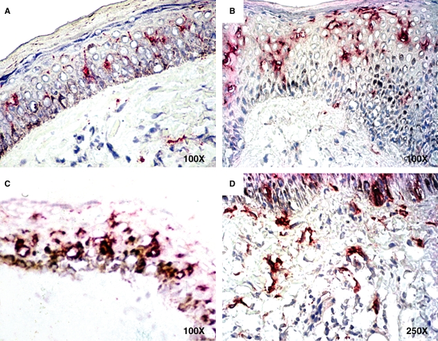
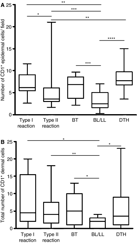
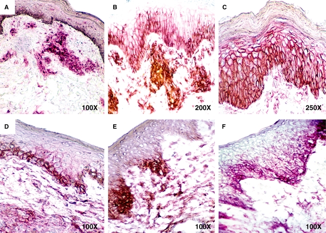
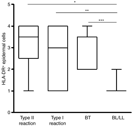
Methods and results: The number and distribution of CD1a+ skin cells and HLA-DR and intercellular adhesion molecule (ICAM)-1 expression were analysed in leprosy skin lesions and in delayed-type hypersensitivity (DTH) tests. The results showed a high number of LCs in tuberculin and lepromin tests, in tuberculoid lesions and in the epidermis and dermis during type I and II reactions. In multibacillary lesions, however, the number of LCs was consistently low in comparison with other groups. Increased numbers of LCs were accompanied by marked HLA-DR and ICAM-1 expression, suggesting a strong relationship between these immunological events.
Conclusions: CD1a+ cells are implicated in the local immunological events taking place after mycobacterial stimuli and may account for the local activation of all types of reactional episodes in leprosy.
Figures
References
-
- Sampaio EP, Sarno EN. Expression and cytokine secretion in the states of immune reactivation in leprosy. Braz. J. Med. Biol. Res. 1998;31:69–76. - PubMed
-
- Sieling PA, Jullien D, Dahlem M, et al. CD1 expression by dendritic cells in human leprosy lesions: correlation with effective host immunity. J. Immunol. 1999;162:1851–1858. - PubMed
-
- Porcelli SA, Modlin RL. CD1 and the expanding universe of T cell antigens. J. Immunol. 1995;155:3709–3710. - PubMed
-
- Zeng Z, Castano AR, Segelke BW, Stura EA, Peterson PA, Wilson IA. Crystal structure of mouse CD1: an MHC-like fold with a large hydrophobic binding groove. Science. 1997;277:339–345. - PubMed
Publication types
MeSH terms
Substances
LinkOut - more resources
Full Text Sources
Research Materials
Miscellaneous
