Review
. 2008 Feb;209(2):313-20.
doi: 10.1016/j.expneurol.2007.08.004. Epub 2007 Aug 22.

Growth factors and combinatorial therapies for CNS regeneration

Affiliations
Review

Growth factors and combinatorial therapies for CNS regeneration

Paul Lu et al. Exp Neurol. 2008 Feb.

Abstract

There has been remarkable progress in the last 20 years in understanding mechanisms that underlie the success of axonal regeneration in the peripheral nervous system, and the failure of axonal regeneration in the central nervous system. Following the identification of these underlying mechanisms, several distinct therapeutic approaches have been tested in in vivo models of spinal cord injury (SCI) to enhance central axonal structural plasticity, including the therapeutic administration of neurotrophic factors. While several tested mechanisms apparently enhance axonal growth, more recent, properly controlled studies indicate that experimental approaches to combine therapies that target distinct neural mechanisms achieve greater axonal growth than therapies applied in isolation. The search for combination therapies that optimize axonal growth after SCI continues.


Figures

Figure. 1
Figure. 1
Spinal cord axon growth is induced by grafts of NGF- or BDNF-secreting cell grafts. (A) Neurofilament immunolabel shows modest axon penetration into control fibroblast graft, whereas (B) NGF-secreting graft is densely penetrated by axons 3 mo post-injury (see Table 1 for list of responding axons). (C) BDA-labeled reticulospinal axons also extensively penetrate a BDNF-secreting bone marrow stromal cell graft (outlined by dashed lines) placed into site of mid-cervical injury. g, graft; h, host; graft shown 3 mo post-injury. (D) Higher magnification of boxed areas from panel C, demonstrating BDA-labeled reticulospinal axons in BDNF-expressing bone marrow stromal cell graft. Scale bar, A-B, 15 μm; C, 210 μm; D, 80 μm.
Figure 2
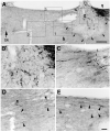
Figure 2
Sensory axons regenerate beyond spinal cord lesion sites after combined administration of intraganglionic cAMP and axonal application of NT-3. (A) Lower magnification view of sagittal section of spinal cord illustrating CTB-labeled dorsal column sensory axons approaching lesion site (arrowhead on upper right), MSC graft in lesion cavity (g; arrows indicate host/graft interface), and region rostral to lesion site (left side of figure). Large arrow and IS indicate rostral Injection Site of NT-3. (B) Boxed area of graft: CTB-labeled sensory axons penetrate graft in lesion site. (C) Higher magnification of box 1 from panel A, demonstrating crossing of CTB-labeled axons from graft into host white matter beyond the graft. Dashed lines indicate host/graft interface. This crossing occurs at a point well away from dorsal or ventral lesion regions, reducing likelihood that axons were spared by lesion. Lesion completeness was confirmed by failure to observe CTB-labeled sensory axons in medulla in lesioned subjects. (D) Higher magnification of box 2 from panel A, demonstrating several varicose CTB-labeled axons extending 0.5 - 0.7 mm beyond lesion site (arrowheads). (E) Higher magnification of box 3 from panel A, showing additional axons extending under the dorsal aspect of the spinal cord beyond the lesion site. Scale bars = 280 μm (A), 44 μm (B-E). (From Lu et al., J Neurosci 2004; 24:6406).

References

    1. Bhatheja K, Field J. Schwann cells: origins and role in axonal maintenance and regeneration. Int J Biochem Cell Biol. 2006;38:1995–1999. - PubMed
    1. Blesch A, Uy HS, Grill RJ, Cheng JG, Patterson PH, Tuszynski MH. Leukemia inhibitory factor augments neurotrophin expression and corticospinal axon growth after adult CNS injury. J Neurosci. 1999;19:3556–3566. - PMC - PubMed
    1. Blits B, Oudega M, Boer GJ, Bunge M, Verhaagen J. Adeno-associated viral vector-mediated neurotrophin gene transfer in the injured adult rat spinal cord improves hindlimb function. Neuroscience. 118:271–281. - PubMed
    1. Boyd JG, Gordon T. Neurotrophic factors and their receptors in axonal regeneration and functional recovery after peripheral nerve injury. Mol Neurobiol. 2003;27:277–324. - PubMed
    1. Bradbury EJ, Khemani S, Von R, King, Priestley JV, McMahon SB. NT-3 promotes growth of lesioned adult rat sensory axons ascending in the dorsal columns of the spinal cord. Eur J Neurosci. 1999;11:3873–3883. - PubMed

Publication types

MeSH terms

Substances