Comparative Study
. 2007 Nov-Dec;128(11-12):601-8.
doi: 10.1016/j.mad.2007.08.006. Epub 2007 Sep 12.

Deletion of Ku80 causes early aging independent of chronic inflammation and Rag-1-induced DSBs

Affiliations
Comparative Study

Deletion of Ku80 causes early aging independent of chronic inflammation and Rag-1-induced DSBs

Valerie B Holcomb et al. Mech Ageing Dev. 2007 Nov-Dec.

Abstract

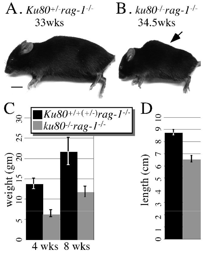
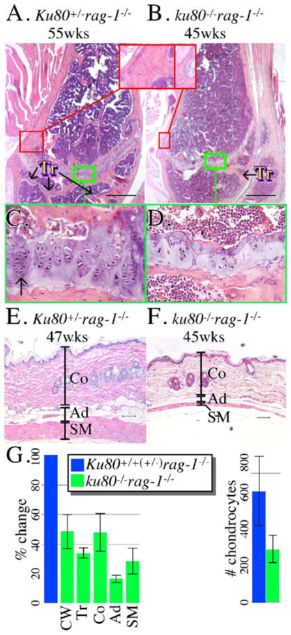
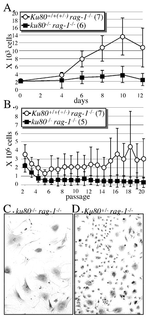
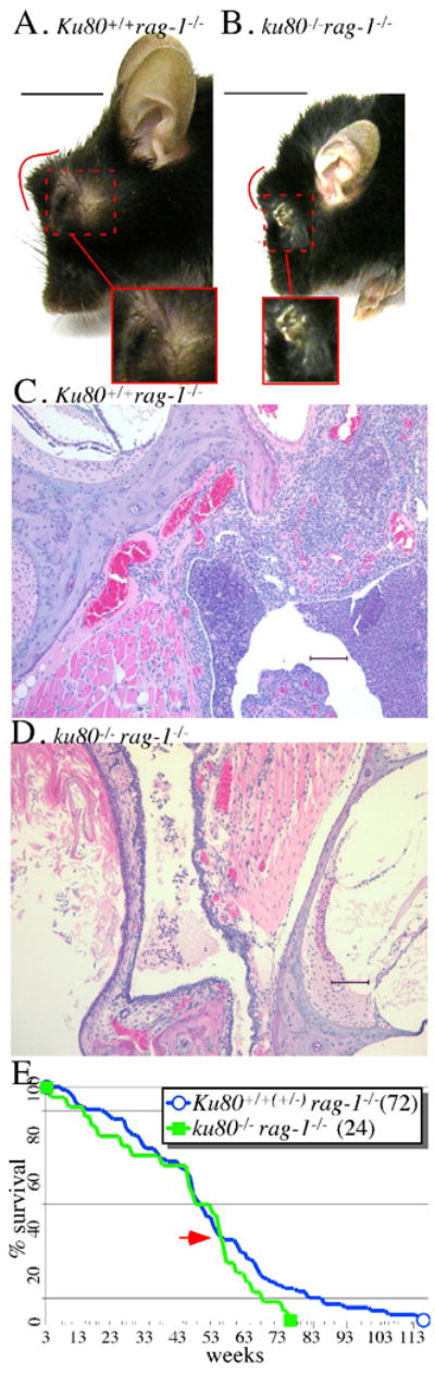
Animal models of premature aging are often defective for DNA repair. Ku80-mutant mice are disabled for nonhomologous end joining; a pathway that repairs both spontaneous DNA double-strand breaks (DSBs) and induced DNA DSBs generated by the action of a complex composed of Rag-1 and Rag-2 (Rag). Rag is essential for inducing DSBs important for assembling V(D)J segments of antigen receptor genes that are required for lymphocyte development. Thus, deletion of either Rag-1 or Ku80 causes severe combined immunodeficiency (scid) leading to chronic inflammation. In addition, Rag-1 induces breaks at non-B DNA structures. Previously we reported Ku80-mutant mice undergo premature aging, yet we do not know the root cause of this phenotype. Early aging may be caused by either defective repair of spontaneous DNA damage, defective repair of Rag-1-induced breaks or chronic inflammation caused by scid. To address this issue, we analyzed aging in control and Ku80-mutant mice deleted for Rag-1 such that both cohorts are scid and suffer from chronic inflammation. We make two observations: (1) chronic inflammation does not cause premature aging in these mice and (2) Ku80-mutant mice exhibit early aging independent of Rag-1. Therefore, this study supports defective repair of spontaneous DNA damage as the root cause of early aging in Ku80-mutant mice.


Figures

Fig. 1
Fig. 1
Gross analysis. Lateral view of a (A) 33 week-old Ku80+/− rag-1−/− and a (B) 34.5 week-old ku80−/− rag-1−/− mouse. Only the ku80−/− rag-1−/− mouse exhibits kyphosis (arrow). Scale: 1 cm. (C) Weight of Ku80+/+(+/−) rag-1−/− (black, 21 mice) and ku80−/− rag-1−/− (gray, 12 mice) mice at 4 and 10 weeks. (D) Total length from eye to base of tail for Ku80+/+(+/−) rag-1−/− (black; 5 mice at 32.8, 36.2, 39.1, 39.1, 40.1 weeks) and ku80−/− rag-1−/− (gray; 5 mice at 19.4, 32.8, 36.2, 40, 40.1 weeks).
Fig. 2
Fig. 2
Microscopic analysis. (A, B) Femur from a (A) 55 week-old Ku80+/− rag-1−/− and a (B) 45 week-old ku80−/− rag-1−/− mouse. Compare thickness of the bone wall (red boxes) and number of trabeculae (Tr). (C, D) Compare the columnar organization and number of chondrocytes from (C) Ku80+/− rag-1−/− and (D) ku80−/− rag-1−/− mouse. Green box from A, B. Arrow indicates chondrocytes. Scale: 500 mm. (E, F) Skin taken from the dorsal–cervico-thoracic region from a (E) 47 week-old Ku80+/− rag-1−/− and a (F) 45 week-old ku80−/− rag-1−/− mouse. Compare thickness of the three skin layers. (Co, Collagen; Ad, Adipose; SM, Skeletal Muscle) Scale: 50 mm. (G) Quantitation of results for Ku80+/+(+/−) rag-1−/− (blue) and ku80−/− rag-1−/− (green) mice. Three control and 3 ku80−/− rag-1−/− mice observed for trabeculae. Four control and 3 ku80−/− rag-1−/− mice observed for cortical wall thickness, skin layers and chondrocytes. Left: % change compared to the mean of the control. Right: the mean number of chondrocytes in the epiphysis. Refer to Experimental procedures for quantification methods.
Fig. 3
Fig. 3
Mouse skin fibroblast analysis. All cells derived from the ears of mice between 32.4–39.5 weeks of age and incubated in atmospheric O2. (A) MSF proliferation curve. Cells (passage 2) derived from seven Rag-1-mutant controls and six double mutant mice. (B) Modified 3T3 analysis to measure lifetime proliferation capacity. MSF (passage 2) are seeded onto two 3.5 cm plates (3.7 × 10⁴ cells/3.5 cm plate). Every 3.5 days cells were trypsinized, combined, counted and seeded at their original concentration onto another 2 plates. As total cell number declined below 7.4 × 10⁴, then cells were seeded onto a single plate at the original concentration. Passaging was discontinued when less than 3.7 × 10⁴ cells remained. (C) Picture of cells (passage 4) derived from the ears of a double mutant mouse. Note the flattened appearance for the double mutant MSF. (D) Picture of cells (passage 4) derived from the ears of a Rag-1-mutant control mouse. Note the spindle-shaped appearance for the Rag-1-mutant control MSF.
Fig. 4
Fig. 4
Opportunistic infections and life span. Suppurative conjunctivitis from a (A) 51-week old Rag-1-mutant control mouse and a (B) 38-week old double mutant mouse. The red line outlines the abnormal shape of the face in profile due to the protruding periocular abscess. The red dashed box shows the abscess adjacent to the eye that has broken through the skin. Scale bar: 1 cm. Otitis media from a (C) 21-week old Rag-1-mutant control mouse and a (D) 16-week old double mutant mouse. Scale bar: 100 mm. (E) Life span of Rag-1-mutant control (blue) and double mutant (green) mice. There is no statistically significant difference between these life spans (log-rank test, Kaplan-Meier survival analysis: p=0.213); however, there is a statistically significant difference when considering the last point their curves meet (red arrow) to the end (p=0.017).

Similar articles

Cited by

References

    1. Baker DJ, Jeganathan KB, Cameron JD, Thompson M, Juneja S, Kopecka A, Kumar R, Jenkins RB, de Groen PC, Roche P, van Deursen JM. BubR1 insufficiency causes early onset of aging-associated phenotypes and infertility in mice. Nat Genet. 2004;36:744–749. - PubMed
    1. Cao L, Li W, Kim S, Brodie SG, Deng CX. Senescence, aging, and malignant transformation mediated by p53 in mice lacking the Brca1 full-length isoform. Genes Dev. 2003;17:201–213. - PMC - PubMed
    1. Chang S, Multani AS, Cabrera NG, Naylor ML, Laud P, Lombard D, Pathak S, Guarente L, DePinho RA. Essential role of limiting telomeres in the pathogenesis of Werner syndrome. Nat Genet. 2004;36:877–882. - PubMed
    1. Chun JJ, Schatz DG, Oettinger MA, Jaenisch R, Baltimore D. The recombination activating gene-1 (RAG-1) transcript is present in the murine central nervous system. Cell. 1991;64:189–200. - PubMed
    1. de Boer J, Andressoo JO, de Wit J, Huijmans J, Beems RB, van Steeg H, Weeda G, van der Horst GT, van Leeuwen W, Themmen AP, et al. Premature aging in mice deficient in DNA repair and transcription. Science. 2002;296:1276–1279. - PubMed

Publication types

MeSH terms