Review
. 2007 Oct;19(5):495-507.
doi: 10.1016/j.ceb.2007.08.002. Epub 2007 Oct 24.

Structure and mechanics of integrin-based cell adhesion

Affiliations
Review

Structure and mechanics of integrin-based cell adhesion

M Amin Arnaout et al. Curr Opin Cell Biol. 2007 Oct.

Abstract

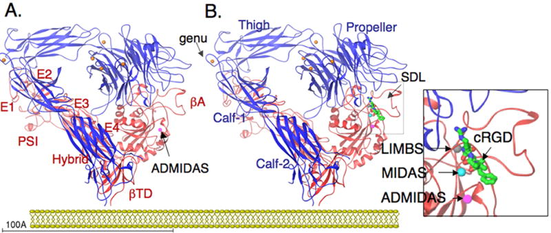
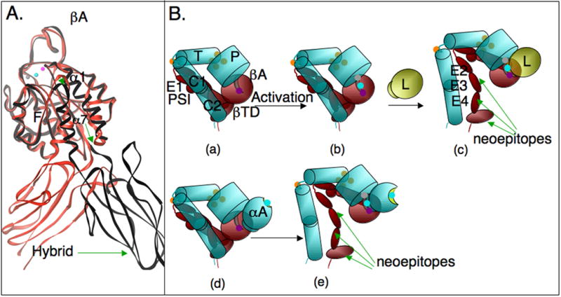
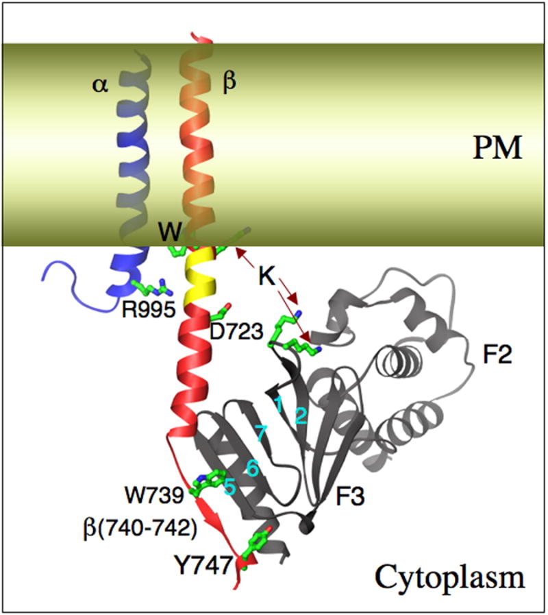
Integrins are alpha/beta heterodimeric adhesion glycoprotein receptors that regulate a wide variety of dynamic cellular processes such as cell migration, phagocytosis, and growth and development. X-ray crystallography of the integrin ectodomain revealed its modular architecture and defined its metal-dependent interaction with extracellular ligands. This interaction is regulated from inside the cell (inside-out activation), through the short cytoplasmic alpha and beta integrin tails, which also mediate biochemical and mechanical signals transmitted to the cytoskeleton by the ligand-occupied integrins, effecting major changes in cell shape, behavior, and fate. Recent advances in the structural elucidation of integrins and integrin-binding cytoskeleton proteins are the subjects of this review.


Figures

Figure 1
Figure 1. Crystal Structure of the integrin 〈V®3 ectodomain
(A) Ribbon diagram of the structure of the unliganded αVβ3 [13]. The protein is bent at a flexible region (genu, arrow), occupied by a metal ion (orange sphere). Four metal ions occupy the base of the propeller (indicated in orange) and an ADMIDAS ion is found in bA (purple sphere). EGF1 and EGF2 are not visible in the structure; their approximate location is indicated. (B) Ribbon diagram of the same structure in complex with the high affinity ligand cRGD [14]; peptide is shown as a ball-and-stick model, with carbons in green, amides in blue and oxygens in red. The specificity-determining loop (SDL) is indicated. Two extra metal ions occupy MIDAS and LIMBS (indicated in the insert). The ectodomain lacks TM segments. Its orientation with respect to a hypothetical membrane approximates the CyoEM structure of full-length aIIbb3 reported by Adair and Yager [31].
Figure 2
Figure 2. Conformational regulation in integrins
(A) Conformational differences in two cRGD-bound ®A domains, one (in red) taken from the cRGD-bound 〈V®3 ectodomain (shown in Figure 1B), and the second (in black) from structure of legless αIIb®3 containing the PSI/Hybrid/®A and propeller domains [19]. cRGD is not shown, but the metal ion triplet (ADMIDAS, MIDAS and LIMBS) indicative of the high affinity state are superimposible, consistent with the absence of differences in the 3 surface loops that form the ligand-binding site. The 〈1 helix straightens in association with a further restructuring of the F/α7 loop (arrow head), a one-turn downward movement 〈7 and a large swing-out of the free hybrid domain (green arrows). (B) Conformational effects of the 〈A domain in 〈A-containing integrins on activation. Inside out activation is much more likely to induce expression of extension/swing-sensitive neoepitopes in the absence of extrinsic ligand in 〈A-containing integrins, which contain their own endogenous ligand in 〈A (ligand-relay hypothesis, see text). Inside-out activated 〈A-lacking integrins generally require exogenous ligand to display such epitopes. The common domains in 〈A-lacking- (a-c) and containing (d-e) integrins are schematically reproduced to scale from the ribbon diagrams shown in Figure 1. P, propeller domain; T, Thigh domain, C1,C2, Calf domains 1 and 2; EGF-like domains 1-4 are labeled, E1-4. The 〈A domain in the lower panel is a schematic, in which the inactive 〈A (with a shallow MIDAS occupied by a metal ion, cyan sphere, d) is not engaged with low affinity ®A (depicted with an ADMIDAS only metal ions, purple, d). Activation (depicted by the triple metal in ®A, b, e) rapidly engages the covalently bound 〈A ligand, exposing hidden eiptopes (e), the nature of which depends on the degree of ligand-induced extension (deadbolt model). Exogenous ligand (L, yellow sphere) would be required to produce similar changes in αA-lacking integrins (b, c). These differences likely impact dynamic interaction under flow.
Figure 3
Figure 3. Integrin activation by talin
The talin F2 and F3 domains, the latter in complex with an integrin ®3 cytoplasmic tail peptide [42]. Key interactions observed or proposed are shown (see text fro details). F3 engages the NPxY-containing membrane distal part of β3-tail (in red), which becomes ordered. A second interaction with the cytosolic membrane-proximal helix disrupts the stabilizing R995-D723 salt-bridge (in inactive integrins), pulls an intramembranous segment (indicated in yellow) into the cytoplasm, aided perhaps by proposed electrostatic interactions between F3 and the acidic lipid head groups in the lipid bilayer (in yellow). The net result is a shortening of the TM segment, which likely disrupts intramembranous α/β packing, leading to activation of the ectodomain.

Similar articles

Cited by

References

    1. Hynes RO. Integrins: a family of cell surface receptors. Cell. 1987;48:549–554. - PubMed
    1. Geiger B, Bershadsky A, Pankov R, Yamada KM. Transmembrane crosstalk between the extracellular matrix--cytoskeleton crosstalk. Nat Rev Mol Cell Biol. 2001;2:793–805. - PubMed
    1. Zaidel-Bar R, Ballestrem C, Kam Z, Geiger B. Early molecular events in the assembly of matrix adhesions at the leading edge of migrating cells. J Cell Sci. 2003;116:4605–4613. - PubMed
    1. Vogel V, Sheetz M. Local force and geometry sensing regulate cell functions. Nat Rev Mol Cell Biol. 2006;7:265–275. - PubMed
    1. Michishita M, Videm V, Arnaout MA. A novel divalent cation-binding site in the A domain of the beta 2 integrin CR3 (CD11b/CD18) is essential for ligand binding. Cell. 1993;72:857–867. - PubMed

Publication types