. 2007 Dec;81(24):13578-86.
doi: 10.1128/JVI.01663-07. Epub 2007 Oct 10.

X box binding protein XBP-1s transactivates the Kaposi's sarcoma-associated herpesvirus (KSHV) ORF50 promoter, linking plasma cell differentiation to KSHV reactivation from latency

Affiliations

X box binding protein XBP-1s transactivates the Kaposi's sarcoma-associated herpesvirus (KSHV) ORF50 promoter, linking plasma cell differentiation to KSHV reactivation from latency

Sam J Wilson et al. J Virol. 2007 Dec.

Abstract

Reactivation of lytic replication from viral latency is a defining property of all herpesviruses. Despite this, the authentic physiological cues for the latent-lytic switch are unclear. Such cues should ensure that viral lytic replication occurs under physiological conditions, predominantly in sites which facilitate transmission to permissive uninfected cells and new susceptible hosts. Kaposi's sarcoma-associated herpesvirus (KSHV) is associated with the B-cell neoplasm primary effusion lymphoma (PEL), in which the virus remains latent. We have previously shown that PEL cells have the gene expression profile and immunophenotype of cycling preplasma cells (plasmablasts). Here, we show that the highly active spliced isoform of plasma cell transcription factor X box binding protein 1 (XBP-1s) is a lytic switch for KSHV. XBP-1s is normally absent in PEL, but the induction of endoplasmic reticulum stress leads to XBP-1s generation, plasma cell-like differentiation, and lytic reactivation of KSHV. XBP-1s binds to and activates the KSHV immediate-early gene ORF50 and synergizes with the ORF50 gene product RTA to induce a full lytic cycle. These data suggest that KSHV remains latent until B-cell terminal differentiation into plasma cells, the transcriptional environment of which provides the physiological "lytic switch" through XBP-1s. This links B-cell terminal differentiation to KSHV lytic reactivation.


Figures

FIG. 1.
FIG. 1.
XBP-1 is unspliced in PEL, but splicing is induced by ER stress. (A) RT-PCR amplification across the XBP-1 intron produces a 249-base-pair amplicon from XBP-1u mRNA and a 223-base-pair amplicon from XBP-1s mRNA. PstI digests only the XBP-1u amplicon. RT-PCR amplification from the Burkitt's lymphoma cell line Raji, the PEL cell lines BC-3, BCBL-1, JSC-1, and HBL-6, and the multiple myeloma cell line SK-MM-2 mRNA shows that XBP-1s is produced in SK-MM-2 cells. In these cells, a slower-migrating, non-PstI-digestible PCR hybrid between XBP-1s and XBP-1u products is visible, similar to hybrids described previously (37). (B) RT-PCR amplification from BC-3 or JSC-1 cells mRNA cultured normally or treated for 1 h with 2 mM DTT. XBP-1s and XBP-1u controls were amplified from pXBPsIG and pXBPuIG, respectively. Composite images of the same gel are shown.
FIG. 2.
FIG. 2.
ER stress induces KSHV lytic replication in an XBP-1-dependent manner. (A) 293T cells harboring recombinant KSHV rKSHV.219 (293T r219-9 cells) were stimulated to induce the KSHV lytic cycle by sodium butyrate or 4 mM DTT treatment overnight. At 48 h posttreatment, cells were analyzed using confocal microscopy. Typical fields singly excited with 488-nm light to detect cells expressing EGFP from the EF1α promoter (green) or 568 nm light to detect cells expressing DsRed from the KSHV lytic cycle PAN promoter (red) are shown magnified at ×60. 293T r219-9 cells were also transduced with lentiviral vectors expressing shRNA XBP-1 or shRNA GFP and treated with DTT as described above. (B) RT-PCR amplification for detection of XBP-1 splicing from 293T r219-9 cell mRNA untreated or treated with DTT.
FIG. 3.
FIG. 3.
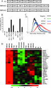
XBP-1 expression in PEL cells induces KSHV lytic replication and expands the secretory apparatus mimicking plasma cell differentiation. (A) Schematic representation of the lentiviral vector genomes used for the expression of XBP-1s and XBP-1u. LTR, long terminal repeat; SFFV, spleen focus-forming virus long terminal repeat promoter; IRES, internal ribosome entry site; WPRE, woodchuck hepatitis virus posttranscriptional regulatory element; 3′SIN LTR, 3′ self-inactivating long terminal repeat. (B) Percentages of RTA-positive PEL cells at 48 h posttransduction with the vectors described above or following TPA treatment. Error bars represent standard errors for triplicate experiments. Percentages were generated by two-parameter flow cytometric analysis. (C) Side scatter histograms generated by flow cytometry of JSC-1 cells at 48 h posttransduction with the vectors described above. (D) Hierarchical clustering of the normalized expression ratios of a subset of host genes. Labeled cDNA from JSC-1 cells that were untreated, TPA treated (TPA), or transduced with the lentiviral vectors described for panel A was hybridized to microarrays in triplicate. Each column represents one condition and each row one gene. Gene expression is shown as a pseudocolored representation of log2 expression ratios with red being above and green below the row/column median level of expression (set to 0), as shown by the scale. Asterisks denote genes which are expressed more highly in plasma cells than in B cells (35).
FIG. 4.
FIG. 4.
XBP-1s expression results in reactivation and recombinant KSHV production. (A) Hierarchical clustering of the normalized expression ratios for the KSHV genes (113 probes) from the microarrays as described in the legend to Fig. 3. (B) Cell clots of JSC-1 cells transduced with lentiviral vectors as described in the legend to Fig. 3A were fixed, paraffin embedded, and stained for lytic KbZIP expression visualized using DAB (brown) counterstained with hematoxylin (blue). (C) HEK 293T BAC36-6 cells (1 × 10⁷; harboring KSHV BAC36) were transfected with the plasmid pXBPsIG and molar equivalents of pIG, pXBPuIG, or pCMV-RTA. At 72 h posttransfection, virus release was quantified by infecting HEK 293Ts and monitoring infection after 48 h by flow cytometry. KSHV infectious units (i.u) are defined as the number of green fluorescent cells (KSHV BAC36 infected) per 2 × 10⁵ cells infected with 1 ml of virus-containing supernatant.
FIG. 5.
FIG. 5.
XBP-1s specifically associates with the KSHV ORF50 promoter, activates ORF50 expression, and synergizes with KSHV RTA in this activity. (A) A schematic representation of the ORF50 promoter used to construct the deletion series of the luciferase reporter constructs. Nucleotide numbers are relative to the KSHV genome sequence NC 003409. (B) HEK 293T cells were transfected with a luciferase reporter plasmid in addition to either pIG (control), pXBPuIG (XBP-1u), or pXBPIG (XBP-1s). At 48 h posttransfection, cells were harvested and luciferase activity was recorded and plotted as a percentage of the activity of the full-length promoter. (C) Chromatin precipitation of XBP-1s. BCBL-1 cells were transfected with pIG or pXBPsIG (which encodes His-tagged XBP-1s). At 48 h posttransfection, cell lysates were immunoprecipitated and PCR amplifications of the KSHV ORF50 promoter, ORF73 promoter, and human DNAJB9 promoter from input, no-antibody controls (No Ab), or immunoprecipitates with anti-HIS (His Ab) were performed. (D) A schematic representation of the ORF50 promoter, including the ORF50 intron, used to construct the deletion series of the DsRed reporter constructs. (E) HEK 293T cells were transfected with a DsRed reporter plasmid in addition to pXBPuIG (XBP-1u) or pXBPIG (XBP-1s), and the percentage of DsRed-express-positive cells was quantified at 48 h posttransfection by flow cytometry. (F) HEK 293T cells were transfected with the full-length ORF50 promoter DsRed reporter plasmid (p50Redi) and pXBPsIG or the molar equivalents of pIG, pXBPuIG, or pCMV-RTA. Half the amount of pXBPsIG, pXBPuIG, pIG, or pCMV-RTA was used in dual transcription factor transfections. The percentages of DsRed-express-positive cells were quantified as described for panel C and are plotted as changes (n-fold) in promoter activity.

Similar articles

Cited by

References

    1. An, J., Y. Sun, M. Fisher, and M. B. Rettig. 2004. Antitumor effects of bortezomib (PS-341) on primary effusion lymphomas. Leukemia 18:1699-1704. - PubMed
    1. Bertolotti, A., Y. Zhang, L. M. Hendershot, H. P. Harding, and D. Ron. 2000. Dynamic interaction of BiP and ER stress transducers in the unfolded-protein response. Nat. Cell Biol. 2:326-332. - PubMed
    1. Besnier, C., Y. Takeuchi, and G. Towers. 2002. Restriction of lentivirus in monkeys. Proc. Natl. Acad. Sci. USA 99:11920-11925. - PMC - PubMed
    1. Bhende, P. M., S. J. Dickerson, X. Sun, W. H. Feng, and S. C. Kenney. 2007. X-box-binding protein 1 activates lytic Epstein-Barr virus gene expression in combination with protein kinase D. J. Virol. 81:7363-7370. - PMC - PubMed
    1. Calfon, M., H. Zeng, F. Urano, J. H. Till, S. R. Hubbard, H. P. Harding, S. G. Clark, and D. Ron. 2002. IRE1 couples endoplasmic reticulum load to secretory capacity by processing the XBP-1 mRNA. Nature 415:92-96. - PubMed

Publication types

MeSH terms

Associated data