. 2008 Jan;82(1):237-45.
doi: 10.1128/JVI.01342-07. Epub 2007 Oct 10.

Double-labeled rabies virus: live tracking of enveloped virus transport

Affiliations

Double-labeled rabies virus: live tracking of enveloped virus transport

Yvonne Klingen et al. J Virol. 2008 Jan.

Abstract

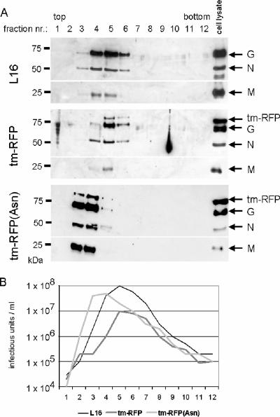
Here we describe a strategy to fluorescently label the envelope of rabies virus (RV), of the Rhabdoviridae family, in order to track the transport of single enveloped viruses in living cells. Red fluorescent proteins (tm-RFP) were engineered to comprise the N-terminal signal sequence and C-terminal transmembrane spanning and cytoplasmic domain sequences of the RV glycoprotein (G). Two variants of tm-RFP were transported to and anchored in the cell surface membrane, independent of glycosylation. As shown by confocal microscopy, tm-RFP colocalized at the cell surface with the RV matrix and G protein and was incorporated into G gene-deficient virus particles. Recombinant RV expressing the membrane-anchored tm-RFP in addition to G yielded infectious viruses with mosaic envelopes containing both tm-RFP and G. Viable double-labeled virus particles comprising a red fluorescent envelope and a green fluorescent ribonucleoprotein were generated by expressing in addition an enhanced green fluorescent protein-phosphoprotein fusion construct (S. Finke, K. Brzozka, and K. K. Conzelmann, J. Virol. 78:12333-12343, 2004). Individual enveloped virus particles were observed under live cell conditions as extracellular particles and inside endosomal vesicles. Importantly, double-labeled RVs were transported in the retrograde direction over long distances in neurites of in vitro-differentiated NS20Y neuroblastoma cells. This indicates that the typical retrograde axonal transport of RV to the central nervous system involves neuronal transport vesicles in which complete enveloped RV particles are carried as a cargo.


Figures

FIG. 1.
FIG. 1.
(A) Schematic presentation of chimeric RFP/RV G constructs. To ensure cell surface expression and incorporation into RV particles, the RFP derivative tdtomato (40) was N-terminally fused to the signal peptide (black boxes) of RV G and C-terminally fused to a C-terminal part of RV G comprising the intracellular cytoplasmic domain, the hydrophobic transmembrane domain (tm), and a membrane-proximal part of the RV G ectodomain (hatched boxes). Predicted glycosylation sites are indicated by diamonds. tm-RFP, no glycosylation site; tm-RFP(Asn), a synthetic glycosylation site was inserted between the signal peptide and RFP; tm-RFP(Asn319), RV ectodomain to aa 316 comprising one authentic glycosylation site (Asn319). (B) Surface expression of RFP/RV G chimeras. Plasmids encoding the indicated RFP constructs under the control of the cytomegalovirus promoter were transfected into BSR T7/5 and neuroblastoma (NA) cells, and 24 h later, RFP autofluorescence in PFA-fixed cells was observed by confocal laser scanning microscopy.
FIG. 2.
FIG. 2.
tm-RFP colocalizes with RV envelope proteins G and M. tm-RFP and RV proteins G and M were expressed in BSR T7/5 cells from transfected cDNAs. After 24 h of incubation, the cells were fixed and permeabilized for immunostainings against RV G or M proteins. (A) After transfection of RV G cDNA only, no tm-RFP autofluorescence was detectable. (B) After transfection of tm-RFP cDNA only, no G-specific immunostaining was detectable. (C) Colocalization of RV G protein and tm-RFP mainly at the cytoplasmic membrane. (D) Colocalization of RV M protein with tm-RFP at the cytoplasmic membrane. (E) After transfection of RV M cDNA only, no G-specific autofluorescence was detectable. All images were acquired at identical microscope settings.
FIG. 3.
FIG. 3.
Incorporation of tm-RFP into SAD ΔG virions. A cell line constitutively expressing tm-RFP was infected with G gene-deleted SAD ΔG. After 2 days of infection, the cell culture supernatants were collected and virions were purified by density gradient centrifugation. Purified virions were mixed with virions from SAD eGFP-P-infected cells, which are known to produce green fluorescent virions (8). After immobilization of virus particles to glass coverslips, red fluorescent particles were detected by laser scanning microscopy. The red and green fluorescent particles were similar in size but were clearly distinguishable by fluorescence from eGFP-labeled virions. Bar, 1 μm.
FIG. 4.
FIG. 4.
Colocalization of tm-RFP and glycoprotein G in infected cells and in virions. (A) Schematic presentation of recombinant RV expressing the indicated tm-RFP constructs. The different tm-RFPs were inserted into the RV genome as an additional transcription unit between the G and L genes. (B) Surface expression of RFP/RV G chimeras in virus-infected cells. After 24 h of infection of BSR-T7/5 cells with the indicated viruses, the cells were fixed and RV G was immunostained with G-specific monoclonal antibody E559. RV G protein and RFP fluorescence colocalized at the cytoplasmic membrane in SAD tm-RFP- and in SAD tm-RFP(Asn)-infected cells. In contrast, no colocalization was detectable in SAD tm-RFP(Asn319)-infected cells. (C) Incorporation of tm-RFP into RV particles. Density gradient-purified supernatant virions were immobilized on glass coverslips, and after fixation with 3% PFA, the virions were immunostained with G-specific antibody E559. Virus particles were detected by confocal laser scanning microscopy.
FIG. 5.
FIG. 5.
Incorporation of RV G and tm-RFP into virions. (A) Density gradient-purified supernatant virions were analyzed by Western blotting using the RV N, M, and G protein-specific antisera. tm-RFP was detected through a G-specific antibody which recognizes the transmembrane domain of RV G protein. The specificity of the tm-RFP signal was also tested with an antibody recognizing DsRed and its derivatives (not shown). (B) The infectious virus titer of each density gradient fraction was determined on BSR T7/5 cells and compared with the virus protein content.
FIG. 6.
FIG. 6.
Growth kinetics of tm-RFP-expressing RVs. The infectious virus titers (IE) in supernatants from virus-infected BSR T7/5 cells (multiplicity of infection = 0.01) were determined at the indicated time points postinfection. In comparison to those of SAD L16, the infectious virus titers of SAD tm-RFP and SAD tm-RFP(Asn) were ∼10-fold decreased.
FIG. 7.
FIG. 7.
Generation of double-labeled RV. (A) Genome organization of control virus SAD L16, SAD tm-RFP, SAD eGFP-P, and the double-labeled SAD eGFP-P tm-RFP. Recombinant SAD eGFP-P tm-RFP virus was generated by combining cDNA plasmids encoding SAD tm-RFP and the previously described SAD eGFP-P. (B) Density gradient-purified supernatant virions from SAD eGFP-P tm-RFP-infected cells were immobilized on glass coverslips. Virus particles were detectable by confocal laser scanning microscopy. (C) Quantification of the fluorescence of a total of 93 eGFP-positive particles. Error bars indicate standard deviations. (D) Double-labeled SAD eGFP-P tm-RFP virions were detectable in endosomes of living BSR T7/5 cells. Confocal fluorescence images with the individual fluorescence channels (top three rows, left and middle panels), a fluorescence merge (top three rows, right panels), and an overlay with nonconfocal DIC images (bottom panels) are shown.
FIG. 8.
FIG. 8.
RV particle movement in axon-like structures of NS20Y cells differentiated by butyryl-cAMP-treatment. The differentiated cells were incubated with double-labeled virus SAD eGFP-P tm-RFP expressing the glycoprotein of the pathogenic RV strain CVS. (A) Attachment of virions to neuronal outgrowths with no or marginal movement. (B) Virus particles which showed slight movement in different directions. (C) Movement of enveloped virus in the anterograde direction (from the top to the bottom). (D) Retrograde transport of double-labeled RV in NS20Y cells at 6 h postinfection. Fluorescent confocal laser scanning microscopy images were merged with DIC images of the neuroblastoma cell. In panel D, the image acquisition was for 218 s at 6.4 s/frame. A video file is provided in the supplemental material.

Similar articles

Cited by

References

    1. Amano, T., E. Richelson, and M. Nirenberg. 1972. Neurotransmitter synthesis by neuroblastoma clones. Proc. Natl. Acad. Sci. USA 69258-263. - PMC - PubMed
    1. Buchholz, U. J., S. Finke, and K. K. Conzelmann. 1999. Generation of bovine respiratory syncytial virus (BRSV) from cDNA: BRSV NS2 is not essential for virus replication in tissue culture, and the human RSV leader region acts as a functional BRSV genome promoter. J. Virol. 73251-259. - PMC - PubMed
    1. Coulon, P., J. P. Ternaux, A. Flamand, and C. Tuffereau. 1998. An avirulent mutant of rabies virus is unable to infect motoneurons in vivo and in vitro. J. Virol. 72273-278. - PMC - PubMed
    1. Dalton, K. P., and J. K. Rose. 2001. Vesicular stomatitis virus glycoprotein containing the entire green fluorescent protein on its cytoplasmic domain is incorporated efficiently into virus particles. Virology 279414-421. - PubMed
    1. Das, S. C., D. Nayak, Y. Zhou, and A. K. Pattnaik. 2006. Visualization of intracellular transport of vesicular stomatitis virus nucleocapsids in living cells. J. Virol. 806368-6377. - PMC - PubMed

Publication types

MeSH terms