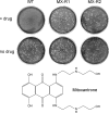
Identification of novel antipoxviral agents: mitoxantrone inhibits vaccinia virus replication by blocking virion assembly
- PMID: 17928345
- PMCID: PMC2168821
- DOI: 10.1128/JVI.00770-07
Identification of novel antipoxviral agents: mitoxantrone inhibits vaccinia virus replication by blocking virion assembly
Abstract
The bioterror threat of a smallpox outbreak in an unvaccinated population has mobilized efforts to develop new antipoxviral agents. By screening a library of known drugs, we identified 13 compounds that inhibited vaccinia virus replication at noncytotoxic doses. The anticancer drug mitoxantrone is unique among the inhibitors identified in that it has no apparent impact on viral gene expression. Rather, it blocks processing of viral structural proteins and assembly of mature progeny virions. The isolation of mitoxantrone-resistant vaccinia strains underscores that a viral protein is the likely target of the drug. Whole-genome sequencing of mitoxantrone-resistant viruses pinpointed missense mutations in the N-terminal domain of vaccinia DNA ligase. Despite its favorable activity in cell culture, mitoxantrone administered intraperitoneally at the maximum tolerated dose failed to protect mice against a lethal intranasal infection with vaccinia virus.
Figures


Similar articles
-
Vaccinia virus DNA ligase recruits cellular topoisomerase II to sites of viral replication and assembly.J Virol. 2008 Jun;82(12):5922-32. doi: 10.1128/JVI.02723-07. Epub 2008 Apr 16. J Virol. 2008. PMID: 18417590 Free PMC article.
-
Mutations in the E9L polymerase gene of cidofovir-resistant vaccinia virus strain WR are associated with the drug resistance phenotype.Antimicrob Agents Chemother. 2006 Dec;50(12):4038-43. doi: 10.1128/AAC.00380-06. Epub 2006 Sep 18. Antimicrob Agents Chemother. 2006. PMID: 16982794 Free PMC article.
-
An orally bioavailable antipoxvirus compound (ST-246) inhibits extracellular virus formation and protects mice from lethal orthopoxvirus Challenge.J Virol. 2005 Oct;79(20):13139-49. doi: 10.1128/JVI.79.20.13139-13149.2005. J Virol. 2005. PMID: 16189015 Free PMC article.
-
From crescent to mature virion: vaccinia virus assembly and maturation.Viruses. 2014 Oct 7;6(10):3787-808. doi: 10.3390/v6103787. Viruses. 2014. PMID: 25296112 Free PMC article. Review.
-
In a nutshell: structure and assembly of the vaccinia virion.Adv Virus Res. 2006;66:31-124. doi: 10.1016/S0065-3527(06)66002-8. Adv Virus Res. 2006. PMID: 16877059 Review.
Cited by
-
A new advanced in silico drug discovery method for novel coronavirus (SARS-CoV-2) with tensor decomposition-based unsupervised feature extraction.PLoS One. 2020 Sep 11;15(9):e0238907. doi: 10.1371/journal.pone.0238907. eCollection 2020. PLoS One. 2020. PMID: 32915876 Free PMC article.
-
Structure-activity studies on the lycorine pharmacophore: A potent inducer of apoptosis in human leukemia cells.Phytochemistry. 2009 May;70(7):913-9. doi: 10.1016/j.phytochem.2009.04.012. Epub 2009 May 20. Phytochemistry. 2009. PMID: 19464034 Free PMC article.
-
A multi-targeting drug design strategy for identifying potent anti-SARS-CoV-2 inhibitors.Acta Pharmacol Sin. 2022 Feb;43(2):483-493. doi: 10.1038/s41401-021-00668-7. Epub 2021 Apr 27. Acta Pharmacol Sin. 2022. PMID: 33907306 Free PMC article.
-
An image-based biosensor assay strategy to screen for modulators of the microRNA 21 biogenesis pathway.Comb Chem High Throughput Screen. 2012 Aug;15(7):529-41. doi: 10.2174/138620712801619131. Comb Chem High Throughput Screen. 2012. PMID: 22540737 Free PMC article.
-
Validation of a high-content screening assay using whole-well imaging of transformed phenotypes.Assay Drug Dev Technol. 2011 Jun;9(3):247-61. doi: 10.1089/adt.2010.0342. Epub 2010 Dec 23. Assay Drug Dev Technol. 2011. PMID: 21182456 Free PMC article.
References
-
- Baldick, C. J., and B. Moss. 1987. Resistance of vaccinia virus to rifampicin conferred by a single nucleotide substitution near the predicted amino terminus of a gene encoding an Mr 62,000 polypeptide. Virology 156:138-145. - PubMed
-
- Borroto-Esoda, K., F. Myrick, J. Feng, J. Jeffrey, and P. Furman. 2004. In vitro combination of amdoxovir and the inosine monophosphate dehydrogenase inhibitors mycophenolic acid and ribavirin demonstrates potent activity against wild-type and drug-resistant variants of human immunodeficiency virus. Antimicrob. Agents Chemother. 48:4387-4394. - PMC - PubMed
-
- Buller, R. M., G. Owens, J. Schriewer, L. Melman, J. R. Beadle, and K. Y. Hostetler. 2004. Efficacy of oral active ether lipid analogs of cidofovir in a lethal mousepox infection. Virology 318:474-481. - PubMed
Publication types
MeSH terms
Substances
Grants and funding
LinkOut - more resources
Full Text Sources
Other Literature Sources

