Review
. 2008 Mar;82(5):2040-55.
doi: 10.1128/JVI.01625-07. Epub 2007 Oct 10.

Viral and host factors in human respiratory syncytial virus pathogenesis

Affiliations
Review

Viral and host factors in human respiratory syncytial virus pathogenesis

Peter L Collins et al. J Virol. 2008 Mar.
No abstract available


Figures

FIG. 1.
FIG. 1.
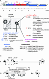
RSV virion, RNA genome, and encoded proteins. (A) The negative-sense RNA genome (strain A2) is depicted 3′ to 5′ showing the extragenic 3′ leader (le) and 5′ trailer (tr) regions and the intervening 10 viral genes (rectangles) that are each expressed as a separate mRNA (21). M2-1 and M2-2 are overlapping open reading frames of the M2 mRNA. The M2 and L genes overlap slightly, and L is expressed by polymerase backtracking (25). (B) Electron photomicrographs showing an RSV virion budding through the plasma membrane of an infected cell (left) and a free virion (right). Protein functions and amino acid lengths (in parentheses, unmodified form) are indicated. (C) Schematic diagrams of the F, G, and SH proteins, with lengths approximately to scale (21). Filled rectangles indicate the hydrophobic cleaved signal sequence (sig.), transmembrane anchors (TM), and fusion peptide (FP). Cross-hatched rectangles indicate heptad repeats (HR) that drive conformational changes involved in fusion. CT, cytoplasmic domain. Potential acceptor sites for N-linked sugars are indicated as downward-facing stalks with an N. For the F protein, the locations and amino acid positions of the two cleavage sites are indicated, as are the cleavage products (F2, p27, F1). For the G protein, the 25 potential acceptor sites for O-linked sugars predicted to be the most likely to be utilized are indicated as downward-facing lollipops. The sequence and disulfide-bonding pattern (dotted lines) of the central domain are shown with the CX3C fractalkine motif boxed and the highly conserved 13-amino-acid sequence of unknown significance underlined. The M-48 translational start site for the secreted form and the mature secreted form is indicated.
FIG. 2.
FIG. 2.
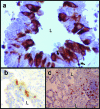
Immunohistochemistry of autopsy lung specimens from a 15-month-old patient with an untreated RSV infection (72). Immunostaining for RSV antigens (a) revealed infection of the bronchiolar epithelium in a near circumferential pattern sparing the basal cells. The basal cells can restore the epithelium, but in the process may lead to mucus metaplasia and remodeling of airways. In addition to infection of the polarized ciliated epithelium, it appears that other cell types, potentially including intraepithelial DCs, can be infected (arrows point to the projections of irregularly shaped RSV-infected cells that are thought to be DCs, based on morphology). Immunostaining for CD1a+ DCs (b) showed that they are not prominent in the lung parenchyma during infection, but when present can have a distinctive morphology with multiple projections that can extend beyond the apical or basal boundary of the columnar epithelium. CD69+ monocytes were the major cell type present in peribronchiolar infiltrates. However, CD3+ T cells also were abundant and, while most of them appeared to be double negative for CD4 and CD8, many were positive for CD8 immunostaining (c). In panels a to c, L indicates airway lumen; in panel c, the lower lumen is occluded with debris and immune cells.
FIG. 3.
FIG. 3.
Estimated contributions of host and viral factors to RSV pathogenesis in the overall pediatric population, as discussed in the text. Factors are placed in the vertical dimension approximately according to the extent to which they are determined by the host (top) or virus (bottom) or a combination (in between). Placement in the horizontal dimension indicates the extent to which they are pathogenic (left) or protective (right). The size of the symbol represents speculated aggregate impact.

Similar articles

Cited by

References

    1. Abu-Harb, M., F. Bell, A. Finn, W. H. Rao, L. Nixon, D. Shale, and M. L. Everard. 1999. IL-8 and neutrophil elastase levels in the respiratory tract of infants with RSV bronchiolitis. Eur. Respir. J. 14139-143. - PubMed
    1. Adkins, B., C. Leclerc, and S. Marshall-Clarke. 2004. Neonatal adaptive immunity comes of age. Nat. Rev. Immunol. 4553-564. - PubMed
    1. Akira, S., S. Uematsu, and O. Takeuchi. 2006. Pathogen recognition and innate immunity. Cell 124783-801. - PubMed
    1. Amanatidou, V., G. Sourvinos, S. Apostolakis, A. Tsilimigaki, and D. A. Spandidos. 2006. T280M variation of the CX3C receptor gene is associated with increased risk for severe respiratory syncytial virus bronchiolitis. Pediatr. Infect. Dis. J. 25410-414. - PubMed
    1. Arnold, R., B. Konig, H. Werchau, and W. Konig. 2004. Respiratory syncytial virus deficient in soluble G protein induced an increased proinflammatory response in human lung epithelial cells. Virology 330384-397. - PubMed

Publication types

MeSH terms