. 2007 Dec;81(24):13325-34.
doi: 10.1128/JVI.01568-07. Epub 2007 Oct 10.

Primary human splenic macrophages, but not T or B cells, are the principal target cells for dengue virus infection in vitro

Affiliations

Primary human splenic macrophages, but not T or B cells, are the principal target cells for dengue virus infection in vitro

Shanley Blackley et al. J Virol. 2007 Dec.

Abstract

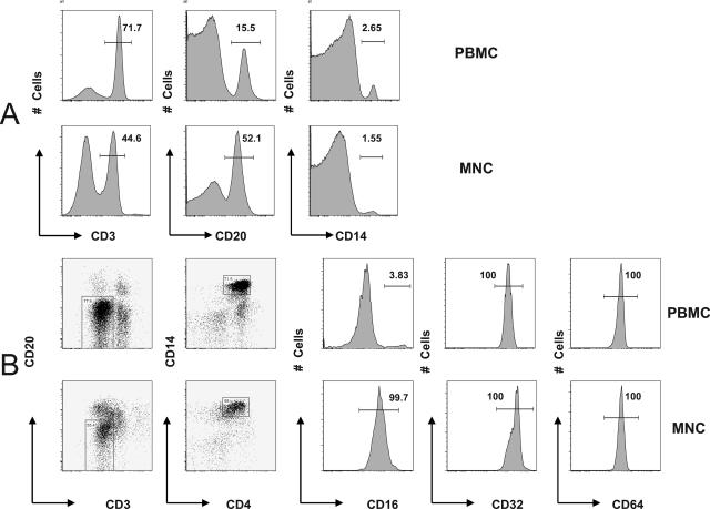
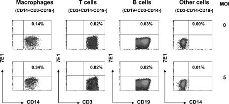
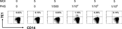
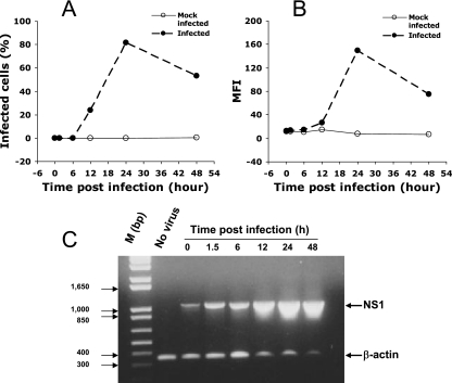
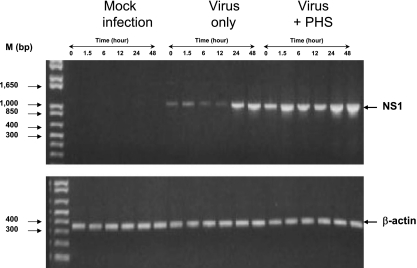
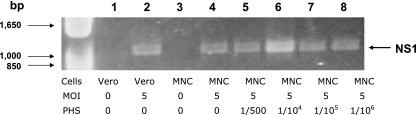
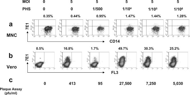
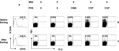
Understanding the pathogenesis of dengue hemorrhagic fever (DHF) and dengue shock syndrome (DSS) requires the precise identification of dengue virus (DV)-permissive target cells. In a previous study using unfractionated human peripheral blood mononuclear cells, we found that monocytes, but not B or T cells, were the principal DV-permissive cells in the absence of DV-immune pooled human sera (PHS) and the major mediators of antibody-dependent enhancement in the presence of PHS. To further identify DV-permissive target cells in other tissues and organs, we isolated human splenic mononuclear cells (MNCs), inoculated them with DV type 2 (strain 16681) in the presence or absence of PHS, and assessed their infection either directly using flow cytometry and reverse transcription-PCR (RT-PCR) assays or indirectly by plaque assay. We found that in the absence of PHS, a small proportion of splenic macrophages appeared to be positive for DV antigens in comparison to staining controls by the flow cytometric assay (0.77% +/- 1.00% versus 0.18% +/- 0.12%; P = 0.07) and that viral RNA was detectable by the RT-PCR assay in MNCs exposed to DV. Additionally, supernatants from cultures of DV-exposed MNCs contained infectious virions that were readily detectable by plaque assay. The magnitude of infection was significantly enhanced in splenic macrophages in the presence of highly diluted PHS (5.41% +/- 3.53% versus 0.77% +/- 1.00%; P = 0.001). In contrast, primary T and B cells were not infected in either the presence or absence of PHS. These results provide evidence, for the first time, that human primary splenic macrophages, rather than B or T cells, are the principal DV-permissive cells in the spleen and that they may be uniquely important in the initial steps of immune enhancement that leads to DHF/DSS in some DV-infected individuals.


Figures

FIG. 1.
FIG. 1.
Comparison of cell subsets between blood PBMCs and splenic MNCs. PBMCs and splenic MNCs were stained with cell subset makers and FcγR markers and compared using FACS analysis as described in Materials and Methods. (A) Distribution of T cells (CD3+), B cells (CD20+), and monocytes/macrophages (CD14+). (B) FcγRI (CD64), FcγRII (CD32), and FcγRIII (CD16) expression in blood PBMCs and splenic MNCs.
FIG. 2.
FIG. 2.
Simultaneous assessment of DV permissiveness among cell subsets. Splenic MNCs were either uninfected (top panel) or infected with DV2-16681 at an MOI of 5 (bottom panel) and stained with anti-E antibody and antibodies to CD3, CD14, and CD19. Results show that only the resident macrophages (CD14+ CD3 CD19) exhibited slightly higher anti-E staining (0.34%) than background staining (0.14%); T cells (CD3+ CD14 CD19), B cells (CD19+ CD3 CD14), and cells that were negative for T-cell, B-cell and monocyte/macrophage markers (CD3 CD14 CD19) exhibited no significant increase in anti-E staining above background.
FIG. 3.
FIG. 3.
Antibody-dependent enhancement of DV infection in splenic macrophages, but not T or B cells. Splenic MNCs were infected by DV2-16681 at an MOI of 5 in the presence or absence of serially diluted PHS (dilutions of 1/500 to 1/106), incubated for 2 days, and then harvested and stained with labeled anti-E antibody as well as antibodies to T cells (CD3), B cells (CD19), or macrophages (CD14); cells were then analyzed using flow cytometry. No infection of T or B cells was detected under any condition. Results show percentages of anti-E antibody staining on macrophages (CD14+ CD3 CD19) for each of the conditions.
FIG. 4.
FIG. 4.
Association between detecting DV RNA and protein. Vero cells were either mock infected or infected by DV2-16681 (MOI of 5); samples were harvested at various time points. The proportion of infected cells (A) and the MFI of infected cells (B) were analyzed by intracellular anti-E antibody staining and FACS analysis. In parallel, viral RNA in infected cells was assessed by RT-PCR for the NS1 gene (C). Results show that infected cells were first detectable at 12 h and peaked at 24 h postinfection; in contrast, mock-infected cells changed little in staining over time (A and B). The relative amount of viral RNA in infected cells remained constant during the first 6 h of infection and increased dramatically from 12 to 48 h postinfection (C). As a control for input cellular RNA, β-actin levels were assessed in the same RT-PCR (C). M, molecular size markers.
FIG. 5.
FIG. 5.
Kinetics of detecting DV gene expression in primary cells. (Top) PBMCs were either mock infected or infected by DV2-16681 (MOI of 5) alone or in the presence of a 1/104 dilution of PHS. Cells were washed and harvested at various times (0, 1.5, 6, 12, 24, and 48 h), and cellular RNAs were extracted for analysis. The presence of the DV NS1 gene product was detected using the RT-PCR assay. A 353-bp β-actin gene fragment was amplified in a separate reaction and used as a control for input cellular RNA. Results show a complete lack of NS1 signal in mock-infected cells and a progressive diminution of NS1 signal during the first 12 h after inoculation with DV alone and its augmentation after 24 to 48 h of infection. After infection by virus-PHS immune complexes, there is a persistence of NS1 during the first 12 h and a late increase NS1 signal at 24 to 48 h postinfection. (Bottom) The amounts of β-actin control are equal in all samples.
FIG. 6.
FIG. 6.
Detection of DV in splenic MNCs in the absence of DV-immune human serum. Splenic MNCs were infected by DV2-16681 at an MOI of 5 in the presence or absence of serially diluted PHS for 2 days. Infection was examined using the RT-PCR assay as described in Materials and Methods. DV-infected or mock-infected Vero cells were used as positive and negative controls, respectively. Results show the detection of the DV NS1 gene in MNCs inoculated with DV alone or in the presence of a range of PHS dilutions (1/500 to 1/106).
FIG. 7.
FIG. 7.
Verification of DV infection using surrogate assays. Splenic MNCs were infected by DV2-16681 at an MOI of 5 in the presence or absence of serially diluted PHS (dilutions of 1/500 to 1/106), incubated for 2 days, and then harvested and stained with labeled anti-E antibody as well as antibodies to T cells (CD3), B cells (CD19), or macrophages (CD14); cells were then analyzed using flow cytometry (a). Culture supernatants were harvested and used to infect Vero cells for three days, and viral infection was assessed using two assays: FACS assay with anti-E antibody staining (b) and plaque assay by immunostaining (c) as described in Materials and Methods. All three methods showed that splenic MNCs were permissive to DV even in the absence of PHS and that the infection was greatly enhanced in the presence of highly diluted PHS.
FIG. 8.
FIG. 8.
Splenic macrophages are a major source of DV production. Splenic MNCs (sample OL3026) were either depleted of CD14+ macrophages or not as described in our previous study (23) and then inoculated with DV2-16681 at an MOI of 5 in the presence or absence of different dilutions of PHS (0, 1/500, 1/104, and 1/105). Supernatant from each of the experimental conditions was harvested 2 days postinfection and used to inoculate Vero cells. After 2 days, Vero cell infection was assessed by staining with labeled anti-E antibody and then analyzed by FACS analysis. Results show the efficiency of CD14 depletion (a) and Vero cell infection with supernatant harvested from unfractionated MNCs (b) or CD14-depleted MNCs (c) under various experimental conditions.

Similar articles

Cited by

References

    1. Avirutnan, P., P. Malasit, B. Seliger, S. Bhakdi, and M. Husmann. 1998. Dengue virus infection of human endothelial cells leads to chemokine production, complement activation, and apoptosis. J. Immunol. 161:6338-6346. - PubMed
    1. Chen, Y. C., and S. Y. Wang. 2002. Activation of terminally differentiated human monocytes/macrophages by dengue virus: productive infection, hierarchical production of innate cytokines and chemokines, and the synergistic effect of lipopolysaccharide. J. Virol. 76:9877-9887. - PMC - PubMed
    1. Diamond, M. S., D. Edgil, T. G. Roberts, B. Lu, and E. Harris. 2000. Infection of human cells by dengue virus is modulated by different cell types and viral strains. J. Virol. 74:7814-7823. - PMC - PubMed
    1. Gordon, S., and P. R. Taylor. 2005. Monocyte and macrophage heterogeneity. Nat. Rev. Immunol. 5:953-964. - PubMed
    1. Green, S., and A. Rothman. 2006. Immunopathological mechanisms in dengue and dengue hemorrhagic fever. Curr. Opin. Infect. Dis. 19:429-436. - PubMed

Publication types

MeSH terms