. 2008 Jan;29(1):170-5.
doi: 10.3174/ajnr.A0769. Epub 2007 Oct 10.

Coinfection of Japanese encephalitis with neurocysticercosis: an imaging study

Affiliations

Coinfection of Japanese encephalitis with neurocysticercosis: an imaging study

S K Handique et al. AJNR Am J Neuroradiol. 2008 Jan.

Abstract

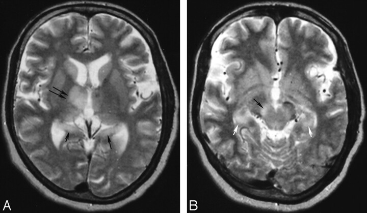
Background and purpose: Coinfection of neurocysticercosis (NCC) and Japanese encephalitis (JE) has been advocated as more than a chance occurrence resulting in poor outcome. We undertook this study to determine whether the association of the 2 infections is more than a chance occurrence, to define the imaging characteristics of coinfections, and to explore the synergistic effect of NCC in JE.

Materials and methods: Sixty-two patients with JE were studied by MR imaging and CT. CT was done in 53 and MR imaging in 53 patients. The diagnosis of JE was established by CSF JE virus immunoglobulin M capture (MAC) enzyme-linked immunosorbent assay (ELISA). NCC was diagnosed from imaging. A control group of 385 patients was evaluated by imaging for prevalence of NCC in the general population.

Results: A significantly high association of NCC with JE (19.3%) was observed in comparison with prevalence of NCC in control subjects (1.04%; P = .0003). JE lesions in coinfection were significantly asymmetric with lateralization to the side of the brain having the maximum NCC or a cyst with edema. The JE lesions in coinfections were more florid, with a significantly higher proportion of abnormal CT scans and more abnormal MR imaging. Coinfections were significantly more common in children. Significantly lower CSF MAC-ELISA units in patients with coinfection reflected low CSF IgM levels, suggesting altered immune status.

Conclusion: In our series, there was a strong association between JE and NCC, and, thus, this coinfection was more than a chance occurrence.


Figures

Fig 1.
Fig 1.
Thirty-one-year-old man with coinfection of JE and NCC (patient no. 2). A and B, Contrast-enhanced CT scans show nonenhancing left thalamic lesion (arrow in A) with a left parietal cysticercus with identifiable scolex and edema (arrow in B). C and D, Axial T2-weighted MR imaging scans done on day 4 of onset of symptoms show a left parietal cysticercus with edema (arrow in C) and T2 hyperintense bilateral thalamic lesions, with predominant lesion on the left side (arrows in D). E, Follow-up axial T2-weighted MR imaging done after 67 days from onset shows resolution of the thalamic lesions.
Fig 2.
Fig 2.
Twelve-year-old girl with coinfection of JE and NCC (patient 5). A, Axial T2-weighted MR imaging done on day 5 after onset of symptoms shows a left frontal cysticercus with T2 hyperintense perifocal edema (arrow). This lesion showed ring enhancement with edema on contrast enhanced CT (not shown). B, Axial T2-weighted MR imaging done on the same day as A shows a left thalamic lesion with mass effect (arrow). The substantia nigra were not involved in this stage (not shown). C and D, Axial T2-weighted MR imaging scans done on day 13 show involvement of both thalami (arrows in C) and both substantia nigra (arrows in D). E, Axial T2-weighted MR imaging done after 45 days of onset shows residual lesions in both substantia nigra (arrows). Residual lesions were also seen in both thalami at this stage (not shown).
Fig 3.
Fig 3.
Thirteen-year-old girl with coinfection of JE and NCC (patient 6). A, Axial T2-weighted MR imaging scan showing a left basal ganglia granulonodular NCC with edema (arrow). B, Axial T2-weighted scan done at the same time as A shows bilateral thalamic involvement, more on the left side (arrows). Note sparing of the basal ganglia in the vicinity of the cyst. C, Axial T2-weighted scan slightly lower than B shows bilateral substantia nigra, left more than right (arrows) and left midbrain tectum (double arrows).
Fig 4.
Fig 4.
Fifty-eight-year-old woman with JE without NCC showing asymmetric lesions. A, Axial T2-weighted MR imaging done on day 3 of onset shows thalamic lesion on the right (double arrows). Note bilateral hippocampal tail involvement (single arrows). B, Axial T2-weighted MR imaging done on same day as A shows right sided substantia nigra (black arrow) and the hippocampal lesions (white arrows). This patient is patient 2 of our previous publication.

References

    1. Kabilan L, Rajendran R, Arunachalam N, et al. Japanese encephalitis in India: an overview. Indian J Paediatr 2004;71:609–15 - PubMed
    1. Kalita J, Misra UK. Comparision of CT scan and MRI findings in the diagnosis of Japanese encephalitis. J Neurol Sci 2000;174:3–8 - PubMed
    1. Shoji H, Kida H, Hino H, et al. Magnetic resonance imaging findings in Japanese encephalitis. White matter lesions. J Neuroimaging 1994;4:206–11 - PubMed
    1. Handique SK, Das RR, Barman K, et al. Temporal lobe involvement in Japanese encephalitis–problems in differential diagnosis. AJNR Am J Neuroradiol 2006;27:1027–31 - PMC - PubMed
    1. Del Brutto OH, Rajshekhar V, White AC Jr, et al. Proposed diagnostic criteria for neurocysticercosis. Neurology 2001;57:177–83 - PMC - PubMed