Comparative Study
. 2007 Oct 10;27(41):10935-46.
doi: 10.1523/JNEUROSCI.1629-07.2007.

Spatial genetic patterning of the embryonic neuroepithelium generates GABAergic interneuron diversity in the adult cortex

Affiliations
Comparative Study

Spatial genetic patterning of the embryonic neuroepithelium generates GABAergic interneuron diversity in the adult cortex

Matthew Fogarty et al. J Neurosci. .

Abstract

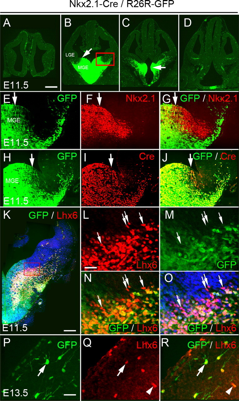
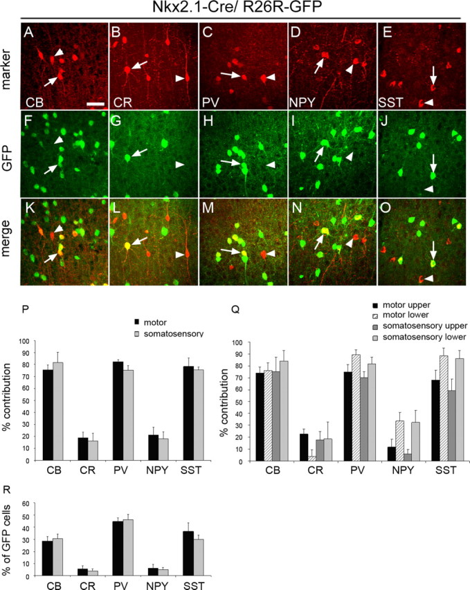
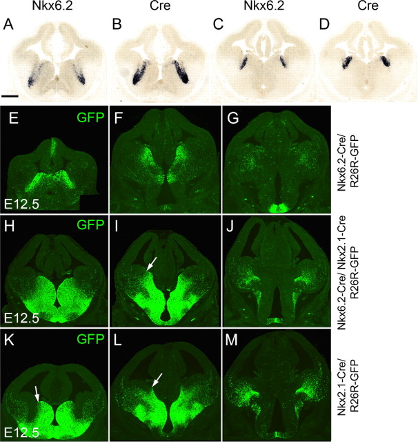
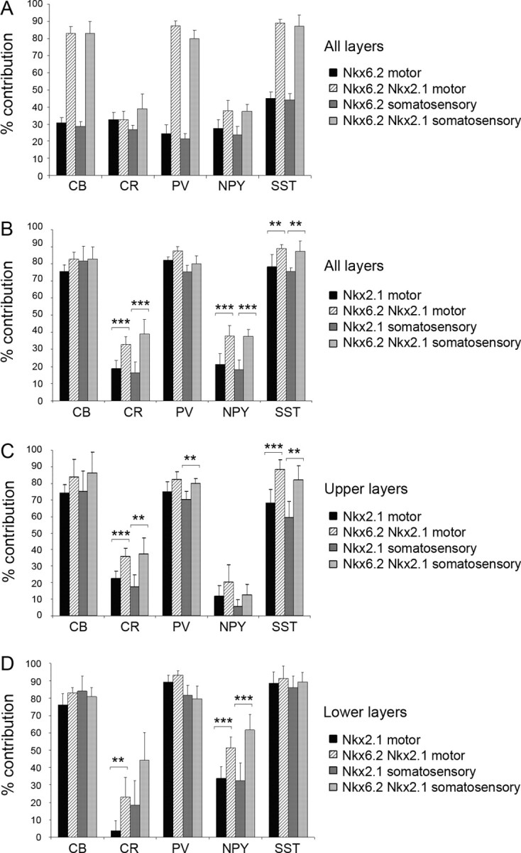
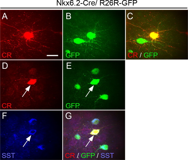
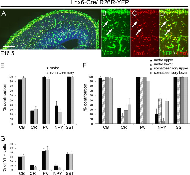
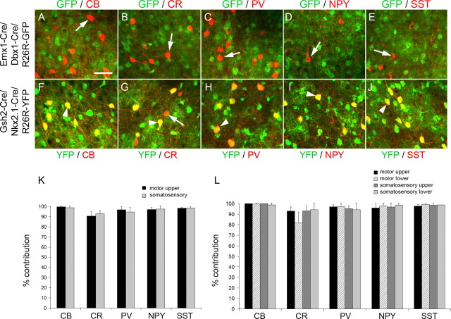
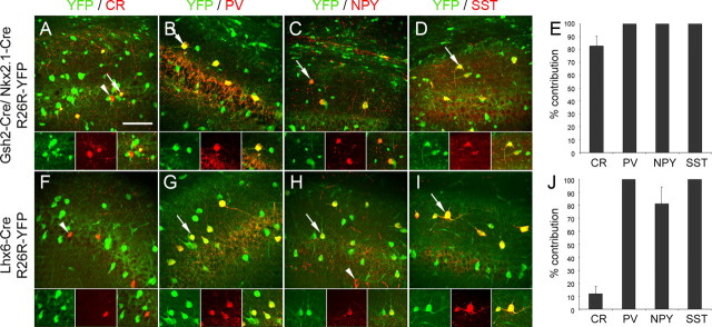
Cortical pyramidal cells are generated from pallial neuroepithelial precursors, whereas GABAergic interneurons originate in subpallial germinal zones and migrate tangentially to reach the cortex. Using Cre-lox technology in transgenic mice and a series of molecular markers that subdivide the subpallial neuroepithelium into small domains, we fate-map precursor pools and identify interneurons generated from each domain. Cortical interneurons expressing calbindin, parvalbumin, and somatostatin are generated exclusively from Lhx6 (Lim homeobox 6)-expressing precursors in the medial ganglionic eminence (MGE). Martinotti cells that coexpress calretinin and somatostatin are generated from the dorsal region of the MGE neuroepithelium that expresses Nkx6.2 (NK2 transcription factor-related 6.2). Most neuropeptide Y-expressing cells and all bipolar calretinin-expressing interneurons are generated outside the MGE, from the germinal zones of the lateral/caudal ganglionic eminences that express Gsh2 (genomic screened homeobox 2). Our data demonstrate that subpallial neuroepithelial domains defined by expression of genetic determinants generate distinct interneuron subtypes, thereby contributing to the generation of cortical interneuron heterogeneity observed in the adult cortex.


Figures

Figure 1.
Figure 1.
Activity of Cre recombinase in Nkx2.1–Cre/R26R–GFP transgenic embryos. A–D, Expression of GFP at four rostrocaudal levels in embryonic day 11.5 (E11.5) transgenic embryos. Arrow in B indicates the dorsal limit of GFP expression. Arrow in C shows expression in the AEP. The red box in B indicates the area shown in E–J. E–G, Comparison between the expression of the endogenous Nkx2.1 gene and GFP at E11.5. Nkx2.1 expression extends farther dorsally than GFP. H–J, Expression of Cre and GFP at E11.5. K–O, Expression of Lhx6 in E11.5 Nkx2.1–Cre/R26R–GFP transgenic embryos. Arrows indicate Lhx6+GFP cells in the dMGE (L–O). P–Q, GFP+ cells in the cortex express Lhx6 (arrows). Arrowheads indicate blood vessels. Scale bars: A–D, 350 μm; E–J, 60 μm; K, 1250 μm; L–O, 25 μm; P–R, 15 μm.
Figure 2.
Figure 2.
Cortical interneurons derived from Nkx2.1-expressing precursors. A–O, Coexpression of GFP with CB, CR, PV, NPY, and SST. Arrows indicate interneurons coexpressing GFP. Arrowheads point to interneurons that are negative for GFP. P, The contribution of Nkx2.1-expressing precursors to interneuron populations expressing CB, CR, PV, NPY, or SST in the motor and somatosensory cortex. The extent of colocalization between GFP and each of the five markers was quantified, and the data are presented as percentage of the total number of cells expressing each of the five markers. Q, The contribution of Nkx2.1-expressing precursors to interneuron populations in the upper and lower layers of the motor and somatosensory cortex. R, The number of Nkx2.1-derived cells coexpressing GFP and one of the markers CB, CR, PV, NPY, or SST are presented as a percentage of the total number of GFP+ cells. Scale bar: (in A) A–O, 20 μm.
Figure 3.
Figure 3.
Activity of Cre recombinase in Nkx6.2–Cre/R26R–GFP and compound Nkx6.2–Cre/Nkx2.1–Cre/R26R–GFP transgenic embryos. A–D, Expression of Nkx6.2 and Cre transcripts in E12.5 Nkx6.2–Cre transgenic embryos. E–G, Expression of GFP at three rostrocaudal levels in E12.5 Nkx6.2–Cre/R26R–GFP transgenic embryos. H–J, Expression of GFP at three rostrocaudal levels in E12.5 Nkx6.2–Cre/Nkx2.1–Cre/R26R–GFP transgenic embryos and comparison with equivalent sections from Nkx2.1–Cre/R26R–GFP transgenic embryos (K–M). Scale bar: (in A) A–M, 300 μm.
Figure 4.
Figure 4.
Cortical interneurons derived from Nkx6.2-, Nkx2.1/Nkx6.2-, and Nkx2.1-expressing precursors. A, The coexpression between GFP and one of the markers CB, CR, PV, NPY, and SST in Nkx6.2–Cre/R26R–GFP and Nkx6.2–Cre/Nkx2.1–Cre/R26R–GFP transgenic mice was quantified and presented as percentage of the total number of cells expressing the markers in the motor and somatosensory cortex. B–D, The contribution of Nkx2.1–Cre-expressing precursors to interneuron populations expressing CB, CR, PV, NPY, and SST was compared with that of Nkx2.1-Cre/Nkx6.2-Cre-expressing precursors (B) in upper (C) and lower (D) levels of the motor and somatosensory cortex. **p < 0.004; ***p < 0.001.
Figure 5.
Figure 5.
Coexpression of CR, SST, and GFP in cortical interneurons of Nkx6.2–Cre/R26R–GFP transgenic mice. A–C, CR+GFP+ cells have a stellate morphology. D–G, Coexpression of SST and GFP in all CR+ cortical interneurons derived from Nkx6.2-expressing precursors. Scale bar, 10 μm.
Figure 6.
Figure 6.
Activity of Cre recombinase in Lhx6–Cre/R26R–YFP transgenic embryos and contribution of Lhx6-expressing precursors to cortical interneuron populations in the adult mouse. A–D, Expression of GFP and Lhx6 in E16.6 transgenic embryos. E, F, The contribution of Lhx6-expressing precursors to interneuron populations expressing CB, CR, PV, NPY, or SST in the motor and somatosensory cortex. The extent of colocalization between YFP and each of the five markers was quantified, and the data are presented as percentage of the total number of cells expressing each of the five markers. G, The number of Lhx6-derived cells coexpressing YFP and one of the markers CB, CR, PV, NPY, or SST are presented as a percentage of the total number of YFP+ cells. Scale bar: A, 100 μm; B–D, 20 μm.
Figure 7.
Figure 7.
Cortical interneurons derived from Emx1/Dbx1- and Gsh2/Nkx2.1-expressing precursors. A–J, Colocalization between GFP/YFP and the interneuron markers CB, CR, PV, NPY, and SST in adult Emx1–Cre/Dbx1–Cre/R26R–GFP transgenic mice (A–E) and Gsh2–Cre/Nkx2.1–Cre/R26R–YFP transgenic mice (F–J). Arrowheads indicate colocalization between GFP/YFP and the interneuron marker. Arrows indicate interneurons that do not express GFP/YFP. K–L, The contribution of Gsh2/Nkx2.1-expressing precursors to interneuron populations expressing CB, CR, PV, NPY, or SST in the motor and somatosensory cortex. The colocalization between GFP and each of the five markers was quantified, and the data are presented as percentage of the total number of cells expressing each of the five markers. Scale bar: (in A) A–J, 20 μm.
Figure 8.
Figure 8.
Interneurons in the hippocampus derived from Gsh2/Nkx2.1 and Lhx6-expressing precursors. A–D, F–I, Colocalization between YFP and the interneuron markers CR, PV, NPY, and SST in CA1 region of the hippocampus in adult Gsh2–Cre/Nkx2.1–Cre/R26R–YFP transgenic mice (A–D) and adult Lhx6–Cre/R26R–YFP transgenic mice (F–I). Arrows indicate colocalization between GFP/YFP and the interneuron marker. Arrowheads indicate interneurons that do not express GFP/YFP. Cells indicated by arrows/arrowheads are shown in the insets. E, J, Quantification of the contribution of Gsh2/Nkx2.1-expressing precursors (E) and Lhx6-expressing precursors (J) to various interneuron populations in the CA1 area of the hippocampus. Data are presented as percentage of the total number of cells expressing each of the four markers. Scale bar: (in A) A–D, F–I, 40 μm.

References

    1. Alifragis P, Liapi A, Parnavelas JG. Lhx6 regulates the migration of cortical interneurons from the ventral telencephalon but does not specify their GABA phenotype. J Neurosci. 2004;24:5643–5648. - PMC - PubMed
    1. Anderson SA, Eisenstat DD, Shi L, Rubenstein JL. Interneuron migration from basal forebrain to neocortex: dependence on Dlx genes. Science. 1997;278:474–476. - PubMed
    1. Anderson SA, Marín O, Horn C, Jennings K, Rubenstein JL. Distinct cortical migrations from the medial and lateral ganglionic eminences. Development. 2001;128:353–363. - PubMed
    1. Awatramani R, Scherer S, Grinspan J, Collarini E, Skoff R, O'Hagan D, Garbern J, Kamholz J. Evidence that the homeodomain protein Gtx is involved in the regulation of oligodendrocyte myelination. J Neurosci. 1997;17:6657–6668. - PMC - PubMed
    1. Bellion A, Wassef M, Metin C. Early differences in axonal outgrowth cell migration GABAergic differentiation properties between the dorsal lateral cortex. Cereb Cortex. 2003;13:203–214. - PubMed

Publication types

Substances