Comparative Study
. 2007 Oct 10;27(41):10993-1002.
doi: 10.1523/JNEUROSCI.2357-07.2007.

Role of nitric oxide in classical conditioning of siphon withdrawal in Aplysia

Affiliations
Comparative Study

Role of nitric oxide in classical conditioning of siphon withdrawal in Aplysia

Igor Antonov et al. J Neurosci. .

Abstract

Nitric oxide (NO) is thought to be involved in several forms of learning in vivo and synaptic plasticity in vitro, but very little is known about the role of NO during physiological forms of plasticity that occur during learning. We addressed that question in a simplified preparation of the Aplysia siphon-withdrawal reflex. We first used in situ hybridization to show that the identified L29 facilitator neurons express NO synthase. Furthermore, exogenous NO produced facilitation of sensory-motor neuron EPSPs, and an inhibitor of NO synthase or an NO scavenger blocked behavioral conditioning. Application of the scavenger to the ganglion or injection into a sensory neuron blocked facilitation of the EPSP and changes in the sensory-neuron membrane properties during conditioning. Injection of the scavenger into the motor neuron reduced facilitation without affecting sensory neuron membrane properties, and injection of an inhibitor of NO synthase had no effect. Postsynaptic injection of an inhibitor of exocytosis had effects similar to injection of the scavenger. However, changes in the shape of the EPSP during conditioning were not consistent with postsynaptic AMPA-like receptor insertion but were mimicked by presynaptic spike broadening. These results suggest that NO makes an important contribution during conditioning and acts directly in both the sensory and motor neurons to affect different processes of facilitation at the synapses between them. In addition, they suggest that NO does not come from either the sensory or motor neurons but rather comes from another source, perhaps the L29 interneurons.


Figures
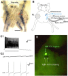
Figure 1.
Figure 1.
L29 facilitatory interneurons express NO synthase. A, NADPH-diaphorase histochemistry labeled three neurons on the left ventral surface of the abdominal ganglion and numerous processes in the neuropil (for details, see Moroz, 2006). R3–R13 and the bag cell clusters are indicated for orientation. Scale bar, 280 μm. B, Approximate locations of the L29 and L30 interneurons, LE sensory neurons, and LFs motor neurons. The siphon nerve and R2 are indicated for orientation. C, Intracellular stimulation of an L29 interneuron recruits a characteristic shower of IPSPs onto itself (C1) attributable to recurrent inhibition from the L30 interneurons (C2). D, Double labeling with intracellular injection of Lucifer yellow and in situ hybridization for Aplysia NOS revealed two NOS-positive L29 interneurons and an NOS-negative L30 interneuron in the same preparation. Intense labeling of the NOS-positive neurons partially masked the Lucifer yellow fluorescence, which was more clearly visible at earlier stages of the NOS staining protocol or at higher magnification.
Figure 2.
Figure 2.
A subset of L29s are NOS negative. A, Double labeling with in situ hybridization for Aplysia NOS (A1) and intracellular injection of Lucifer yellow (A2) revealed that at least two L29s (indicated by 1 and 2) in the same ganglion are NOS positive, but others (e.g., 3) are NOS negative. LE sensory neurons and LFs motor neurons were all NOS negative. Scale bar, 110 μm. B, NOS-positive and NOS-negative L29 interneurons were functionally electrically coupled.
Figure 3.
Figure 3.
NO is involved in facilitation and conditioning. A, Application of the NO donor DEA/NO produces facilitation of the monosynaptic EPSP from an LE sensory neuron to an LFS motor neuron by a direct action in the sensory neuron. A1, Example of the EPSP before (Pretest) and 10 min after (Posttest) application of DEA/NO (10 μm) to the abdominal ganglion. A2, Average results from experiments like the one shown in A1 (n = 7) as well as control experiments without DEA/NO (n = 5) and experiments in which DEA/NO was applied after injection of the NO scavenger oxymyoglobin into either the sensory (n = 6) or motor (n = 6) neuron. There was a significant overall effect of group for the EPSP (F(3,20) = 5.19; p < 0.01) but not for LE membrane resistance (Rm LE). The data have been normalized to the average of the three pretests in each experiment. The average pretest EPSP amplitude was 6.4 mV, not significantly different in the four groups by a one-way ANOVA. Myo, Myoglobin. B, Bathing the abdominal ganglion in inhibitors of NO synthase or PKG blocks conditioning. B1, Experimental preparation. SN, Sensory neuron; MN, motor neuron. B2, Training protocol. For details, see Materials and Methods. B3, Average siphon-withdrawal reflex (SWR) on each test in groups that received paired (P) or unpaired (UP) training with the abdominal ganglion bathed in either normal seawater (n = 36 paired and 36 unpaired), the NO synthase inhibitor Nω-nitro-l-arginine (NOArg; n = 15 and 15), or the PKG inhibitor KT5823 (n = 21 and 21). There was a significant overall drug × pairing interaction (F(2,138) = 6.06; p < 0.01). Responses have been normalized to the value on the pretest in each experiment. The overall average pretest value was 3.0 mm, and the average response to the first tail-shock US was 5.9 mm. These values were not significantly different between the different experimental groups in one-way ANOVAs. In this and subsequent figures, error bars indicate SEMs. *p < 0.05, +p < 0.05, one-tailed for the difference between the experimental and control groups, and #p < 0.05, +p < 0.05, one-tailed for the reduction of that effect by the drug at each test.
Figure 4.
Figure 4.
Neural correlates of conditioning in LE sensory neurons and LFS motor neurons. A, Examples of siphon withdrawal (SW), evoked firing of an LFS siphon motor neuron and an LE siphon sensory neuron, the membrane resistance of each neuron, and the monosynaptic EPSP from the LE neuron to the LFS neuron on the pretest and final posttest after paired or unpaired training with the abdominal ganglion bathed in normal seawater. B, Average results from experiments like the ones shown in A [n = 20 paired (P) and 20 unpaired (UP)]. Results from four different control groups (bathing the abdominal ganglion in normal seawater or inactive myoglobin, or injecting vehicle into an LE sensory neuron or heat-inactivated botulinum toxin into an LFS motor neuron; n = 5 paired and 5 unpaired per group) were not significantly different on any measure and have been pooled. The data have been normalized to the value on the pretest in each experiment. The overall average pretest values in the experiments shown in Figures 4–8 were 1.8 mm for siphon withdrawal, 14.1 spikes for evoked LFS firing, 3.4 spikes for evoked LE firing, and 6.6 mV for the amplitude of the EPSP. The average response to the first tail-shock US was 4.3 mm. These values were not significantly different between the different experimental groups. SWR, Siphon-withdrawal reflex; Rm LE, LE membrane resistance.
Figure 5.
Figure 5.
Bathing the abdominal ganglion in a scavenger of NO, oxymyoglobin, blocks conditioning, facilitation of the EPSP, and the changes in sensory neuron membrane properties. A, Examples with the abdominal ganglion bathed in oxymyoglobin. B, Average results from experiments like the ones shown in A (black symbols; n = 5 and 5) compared with the average control results from Figure 4 (white symbols). SW, Siphon withdrawal; SWR, siphon-withdrawal reflex; Myo, Abd, abdominal ganglion bathed in myoglobin; Rm LE, LE membrane resistance; Con, control; Myo, myoglobin; P, paired; UP, unpaired.
Figure 6.
Figure 6.
Injecting oxymyoglobin into an LE sensory neuron blocks facilitation and the changes in sensory neuron membrane properties. A, Examples after injection of oxymyoglobin into the LE sensory neuron. B, Average results from experiments like the ones shown in A (n = 5 and 5). SW, Siphon withdrawal; SWR, siphon-withdrawal reflex; Myo, LE, LE neuron injected with myoglobin; Rm LE, LE membrane resistance; Con, control; Myo, myoglobin; P, paired; UP, unpaired.
Figure 7.
Figure 7.
Injecting oxymyoglobin into an LFS motor neuron reduces facilitation, and injecting an inhibitor of NO synthase into the motor neuron has no effect. A1, Examples after injection of oxymyoglobin into the LFS motor neuron. A2, Average results from experiments like the ones shown in A1 (n = 5 and 5). B, Average results after injection of Nω-nitro-l-arginine (NOArg) into the LFS neuron (n = 7). SW, Siphon withdrawal; SWR, siphon-withdrawal reflex; Myo, LFS, LFS neuron injected with myoglobin; Rm LE, LE membrane resistance; NOArg, LFS, LFS neuron injected with Nω-nitro-l-arginine; Con, control; Myo, myoglobin; P, paired; UP, unpaired.
Figure 8.
Figure 8.
Injecting botulinum toxin into an LFS motor neuron reduces facilitation. A, Examples after injection of the light chain of botulinum toxin type B into the LFS motor neuron. B, Average results from experiments like the ones shown in A (n = 8 and 6). SW, Siphon withdrawal; SWR, siphon-withdrawal reflex; Botx, LFS, LFS neuron injected with in botulinum toxin; Rm LE, LE membrane resistance; Con, control; Btx, botulinum toxin; P, paired; UP, unpaired.
Figure 9.
Figure 9.
Changes in the EPSP during conditioning are mimicked by presynaptic spike broadening. A, Changes in the peak amplitude of the EPSP and the ratio of the late (75 ms after the peak) and early (peak) parts of the EPSP in each of the experiments shown in Figures 4–8. The data on the posttest have been normalized to the values on the pretest in each experiment. The overall average pretest ratio was 27%. There were no significant differences between the pretest ratios or the slopes of the linear regressions for the different groups. B, Changes in the EPSP during conditioning are mimicked by 4-AP. B1, The experimental protocol for examining the effects of the drugs on the EPSP. B2, Examples of the action potential in an LE sensory neuron and the EPSP in an LFS motor neuron before and after bathing the abdominal ganglion in 4-AP, shown on two timescales to illustrate changes in the EPSP (left) and the action potential (right). B3, Changes in the peak amplitude of the EPSP and the ratio of the late (75 ms after the peak) and early (peak) parts of the EPSP when the ganglion was bathed in APV, CNQX, 4-AP, or 4-AP in ASW with 2× normal Ca2+ (4-AP HiCa) (n = 5, 5, 5, and 4). There were significant effects of drug on both the peak amplitude (F(2,16) = 22.13; p < 0.001) and ratio (F = 10.06; p < 0.01). The average of the three test values has been normalized to the average of the three control (predrug) values in each experiment. The overall average control values were 7.9 mV for the peak amplitude and 39% for the ratio. These values were not significantly different in experiments with high Ca2+. For the 4-AP experiments, the size of the symbols is proportional to the amount of broadening of the presynaptic action potential measured from the peak to 10% of peak on the falling phase. P, Paired; UP, unpaired; Myo, myoglobin; Abd, abdominal ganglion; Botx, botulinum toxin.

References

    1. Abrams TW, Yovell Y, Onyike CU, Cohen JE, Jarrard HE. Analysis of sequence-dependent interactions between calcium and transmitter stimuli in activating adenylyl cyclase in Aplysia: possible contributions to CS-US sequence requirement during conditioning. Learn Mem. 1998;4:496–509. - PubMed
    1. Antonov I, Kandel ER, Hawkins RD. The contribution of facilitation of monosynaptic PSPs to dishabituation and sensitization of the Aplysia siphon withdrawal reflex. J Neurosci. 1999;19:10438–10450. - PMC - PubMed
    1. Antonov I, Antonova I, Kandel ER, Hawkins RD. The contribution of activity-dependent synaptic plasticity to classical conditioning in Aplysia. J Neurosci. 2001;21:6413–6422. - PMC - PubMed
    1. Antonov I, Antonova I, Kandel ER, Hawkins RD. Activity-dependent presynaptic facilitation and Hebbian LTP are both required and interact during classical conditioning in Aplysia. Neuron. 2003;37:135–147. - PubMed
    1. Antonova I, Arancio O, Trillat A-C, Wang H-G, Zablow L, Udo H, Kandel ER, Hawkins RD. Rapid increase in clusters of presynaptic proteins at onset of long-lasting potentiation. Science. 2001;294:1547–1550. - PubMed

Publication types

MeSH terms

LinkOut - more resources