

Serial organization of human behavior in the inferior parietal cortex
- PMID: 17928444
- PMCID: PMC6672846
- DOI: 10.1523/JNEUROSCI.1986-07.2007
Serial organization of human behavior in the inferior parietal cortex
Abstract
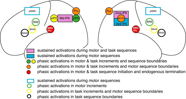
The parietal cortex is involved in a wide range of cognitive functions in humans including associative functions between multiple sensorimotor spaces, attentional control, and working memory. Little is known, however, about the role and the functional organization of the parietal cortex in action planning and sequential cognition. Moreover, the respective contributions of parietal and frontal regions to action planning remains poorly understood. To address this issue, we designed a functional magnetic resonance imaging protocol requiring subjects to perform overlearned sequences of motor acts and sequences of cognitive tasks. The results reveal only a single bilateral region in the cerebral cortex located in the intraparietal sulcus (IPS; Brodmann's area 40) exhibiting sustained activations during the execution of both motor and task sequences. Additional analyses of phasic activations during sequence execution further suggest a functional dissociation between the left IPS, involved in representing and processing the abstract serial structure of ongoing behavioral sequences regardless of their hierarchical structure, and the right IPS, involved in preparing successive sensorimotor sets that compose such behavioral sequences. We show that this parietal system functionally differs from the frontal system that was previously identified as controlling action selection with respect to the hierarchical rather than serial structure of behavioral plans. Thus, our results reveal the central role of the bilateral intraparietal sulcus in high-order sequential cognition and suggest a major functional segregation within the frontoparietal network mediating action planning, with the frontal and parietal sector involved in processing the hierarchical and serial structure of action plans, respectively.
Figures


Similar articles
-
Visual Short-Term Memory Activity in Parietal Lobe Reflects Cognitive Processes beyond Attentional Selection.J Neurosci. 2018 Feb 7;38(6):1511-1519. doi: 10.1523/JNEUROSCI.1716-17.2017. Epub 2018 Jan 8. J Neurosci. 2018. PMID: 29311140 Free PMC article.
-
Short-term memory and the left intraparietal sulcus: focus of attention? Further evidence from a face short-term memory paradigm.Neuroimage. 2007 Mar;35(1):353-67. doi: 10.1016/j.neuroimage.2006.12.008. Epub 2006 Dec 15. Neuroimage. 2007. PMID: 17240164
-
How verbal and spatial manipulation networks contribute to calculation: an fMRI study.Neuropsychologia. 2008;46(9):2403-14. doi: 10.1016/j.neuropsychologia.2008.03.001. Epub 2008 Mar 18. Neuropsychologia. 2008. PMID: 18406434
-
Neurocognitive contributions to motor skill learning: the role of working memory.J Mot Behav. 2012;44(6):445-53. doi: 10.1080/00222895.2012.672348. J Mot Behav. 2012. PMID: 23237467 Free PMC article. Review.
-
Using predictive motor control processes in a cognitive task: behavioral and neuroanatomical perspectives.Adv Exp Med Biol. 2009;629:337-54. doi: 10.1007/978-0-387-77064-2_17. Adv Exp Med Biol. 2009. PMID: 19227508 Free PMC article. Review.
Cited by
-
Compensatory recombination phenomena of neurological functions in central dysphagia patients.Neural Regen Res. 2015 Mar;10(3):490-7. doi: 10.4103/1673-5374.153701. Neural Regen Res. 2015. PMID: 25878601 Free PMC article.
-
The manipulative complexity of Lower Paleolithic stone toolmaking.PLoS One. 2010 Nov 3;5(11):e13718. doi: 10.1371/journal.pone.0013718. PLoS One. 2010. PMID: 21072164 Free PMC article.
-
Grasping neurons of monkey parietal and premotor cortices encode action goals at distinct levels of abstraction during complex action sequences.J Neurosci. 2011 Apr 13;31(15):5876-86. doi: 10.1523/JNEUROSCI.5186-10.2011. J Neurosci. 2011. PMID: 21490229 Free PMC article.
-
Testosterone Modulates Altered Prefrontal Control of Emotional Actions in Psychopathic Offenders(1,2,3).eNeuro. 2016 Feb 8;3(1):ENEURO.0107-15.2016. doi: 10.1523/ENEURO.0107-15.2016. eCollection 2016 Jan-Feb. eNeuro. 2016. PMID: 26878057 Free PMC article.
-
The posterior parietal cortex encodes in parallel both goals for double-reach sequences.J Neurosci. 2008 Oct 1;28(40):10081-9. doi: 10.1523/JNEUROSCI.3423-08.2008. J Neurosci. 2008. PMID: 18829966 Free PMC article.
References
-
- Aboitiz F, Garcia VR. The evolutionary origin of the language areas in the human brain. A neuroanatomical perspective. Brain Res Brain Res Rev. 1997;25:381–396. - PubMed
-
- Andersen RA, Snyder LH, Bradley DC, Xing J. Multimodal representation of space in the posterior parietal cortex and its use in planning movements. Annu Rev Neurosci. 1997;20:303–330. - PubMed
-
- Asari T, Konishi S, Jimura K, Miyashita Y. Multiple components of lateral posterior parietal activation associated with cognitive set shifting. NeuroImage. 2005;26:694–702. - PubMed
-
- Baker SC, Rogers RD, Owen AM, Frith CD, Dolan RJ, Frackowiak RS, Robbins TW. Neural systems engaged by planning: a PET study of the Tower of London task. Neuropsychologia. 1996;34:515–526. - PubMed
-
- Barash S. Paradoxical activities: insight into the relationship of parietal and prefrontal cortices. Trends Neurosci. 2003;26:582–589. - PubMed
Publication types
MeSH terms
LinkOut - more resources
Full Text Sources