Comparative Study
. 2007 Oct 10;27(41):11047-55.
doi: 10.1523/JNEUROSCI.2733-07.2007.

Human immunodeficiency virus-1/surface glycoprotein 120 induces apoptosis through RNA-activated protein kinase signaling in neurons

Affiliations
Comparative Study

Human immunodeficiency virus-1/surface glycoprotein 120 induces apoptosis through RNA-activated protein kinase signaling in neurons

Mehrdad Alirezaei et al. J Neurosci. .

Abstract

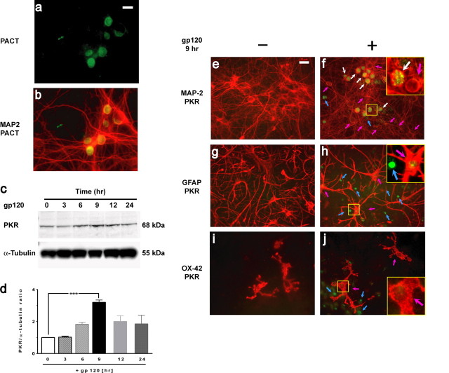
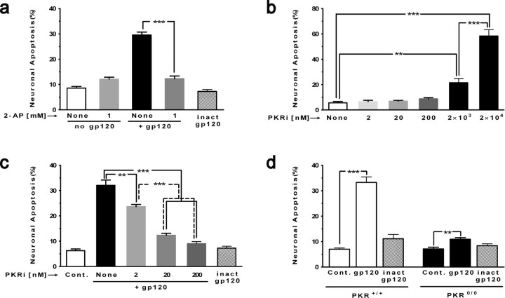
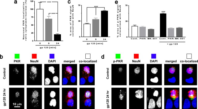
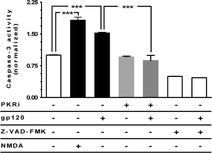
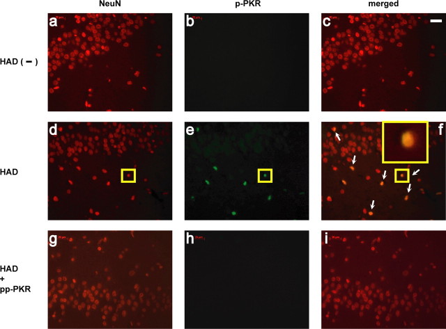
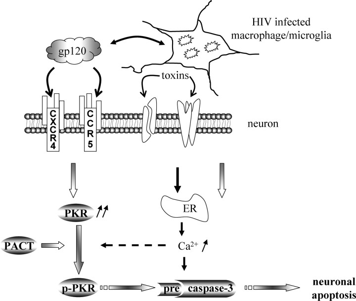
Previous work has demonstrated that the surface glycoprotein (gp120) of human immunodeficiency virus-1 (HIV-1) can induce damage and apoptosis of neurons both in vitro and in vivo. In this report, we provide evidence that double-stranded RNA-activated protein kinase (PKR), a stress kinase, is involved in HIV/gp120-associated neurodegeneration. In cultures of mixed cortical cells, HIV/gp120 increased the protein level of PKR. Additionally, PKR was phosphorylated in neurons but not glia after exposure to gp120. The use of two independent pharmacological inhibitors of PKR activity abrogated neuronal cell death induced by gp120. Cortical neurons from PKR knock-out mice were significantly protected from neurotoxicity induced by gp120, further validating the pivotal proapoptotic function of PKR. gp120-induced phosphorylated PKR localized prominently to neuronal nuclei; PKR inhibition or the NMDA receptor antagonist MK-801 [(+)-5-methyl-10,11-dihydro-5H-dibenzo [a,d] cyclohepten-5,10-imine maleate] abrogated this effect. PKR inactivation also inhibited gp120-induced caspase-3 activation, consistent with its neuroprotective effect. Finally, brain tissue from individuals diagnosed with HIV-associated dementia (HAD), but not HIV infection alone, contained the activated form of PKR, which localized predominantly to neuronal nuclei. Together, these results identify PKR as a critical mediator of gp120 neurotoxicity, suggesting that activation of PKR contributes to the neuronal injury and cell death observed in HAD.


Figures

Figure 1.
Figure 1.
PACT and PKR expression in neurons. a, b, PACT (a; green) and merged image of PACT/MAP-2 (b) expression in neurons in cortical mixed cultures. c, PKR was upregulated in cortical cell cultures exposed to 200 pm gp120 in a time-dependent manner. Western blot analysis was performed on lysates, using α-tubulin as the loading control. d, Ratios between total PKR and α-tubulin were normalized to the value at time 0 h, and mean ± SEM (n = 4) is shown. ***p < 0.001. e, f, Double immunostaining with MAP-2 (red) and PKR (green) antibodies of control cortical cell cultures (untreated) and those exposed to gp120 for 9 h. Upregulated PKR (green) in neurons is indicated with white arrows and in glia with blue arrows. Neurons (red) without PKR induction are indicated with pink arrows. Analysis of 2000 neurons showed that the PKR is upregulated in 36 ± 1.1% of neurons exposed to gp120 for 9 h. g, h, Double immunostaining of cortical cultures exposed to gp120 with GFAP (red) and PKR (green) antibodies. Blue arrows indicate PKR induction in nonastrocytic cells, and pink arrows indicate astrocytes with extremely low PKR expression. i, j, Double immunostaining with OX-42 (specific for microglia, red) and with PKR (green). PKR upregulation in nonmicroglial cells (blue arrows) and microglia without PKR expression are indicated with pink arrows. In the cultures exposed to gp120 (f, h, j), higher-magnification images of the area outlined in yellow are shown in the insets. Scale bars, 20 μm.
Figure 2.
Figure 2.
Prominent role of PKR in gp120 neurotoxicity. When indicated, cortical cell cultures were pretreated with an inhibitor of PKR (30 min) and then exposed to gp120 (200 pm). Immunostaining was performed with an antibody directed against MAP-2 to identify neurons and DAPI for nuclear staining. Apoptotic nuclei were counted from different regions on coverslips. a, 2-AP (1 mm) completely protected neurons against toxicity induced by gp120. 2-AP by itself was not neurotoxic. ***p < 0.001 for n = 4 experiments. b, PKRi was nontoxic at 2, 20, and 200 nm but became toxic at in the micromolar range. ***p < 0.001, **p < 0.01 for n = 3 experiments. c, PKRi (2–200 nm) abrogated gp120 neurotoxicity in a dose-dependent manner. Heat-inactivated gp120 (inact gp120) served as a control (Cont.) and was not neurotoxic. ***p < 0.001, **p < 0.01 for n = 4 experiments. d, Only minimal neurotoxicity occurs in cortical cultures from PKR knock-out mice versus wild type (the phenotypic appearance of PKR0/0 cultures resembled that of wild-type mouse and rat cortical cell cultures; data not shown). Heat-inactivated (inact) gp120 was used as a negative control for the neurotoxicity experiments. ***p < 0.001, **p < 0.01 for n = 4 experiments. All values are mean ± SEM.
Figure 3.
Figure 3.
PKR and p-PKR translocation to nucleus in neurons exposed to gp120. a, PKR colocalization with MAP-2 (cytoplasmic) and DAPI (nuclear) illustrates the PKR translocation to nuclei in cultures exposed to gp120. ***p < 0.001 for n = 4 experiments. b, Images acquired using confocal microscopy of neurons (NeuN positive; red) exposed to gp120 for 24 h with PKR (green) and counterstained with DAPI. Colocalized PKR with NeuN is shown in white in the far right panels. c, PKR colocalization with NeuN revealed an increase of PKR in nuclei after exposure to gp120 for 24 h. ***p < 0.001 for n = 4 experiments. d, Images acquired using confocal microscopy of neurons (NeuN positive, red) exposed to gp120 for 24 h with p-PKR (green) and counterstained with DAPI. Colocalized p-PKR with NeuN is shown in white in the far right panels. e, Increased colocalization of p-PKR and NeuN in neurons exposed to gp120 after 24 h, which is abrogated in the presence of PKRi or MK-801. ***p < 0.001 for n = 4 experiments. Cont., Control. All values are mean ± SEM.
Figure 4.
Figure 4.
PKR inactivation inhibits caspase-3 activity induced by gp120. Cultures were pretreated with 200 nm PKRi and then exposed to gp120 for 18 h. PKRi pretreatment in cortical cultures reduced caspase-3 activity induced by gp120. NMDA (100 μm for 10 min) was used as a positive control for activating caspase-3. Z-VAD-FMK (N-benzyloxycarbonyl-Val-Ala-Asp-fluoromethylketone) (an irreversible inhibitor of caspase-3 activity) was used as a negative control for the assay. ***p < 0.001 for n = 4 experiments. Values are mean ± SEM fold increase in activity.
Figure 5.
Figure 5.
The activated form of PKRi localized in neuronal nuclei from autopsied brain sections of HAD patients. a–c, Hippocampal tissue from an HIV-positive but HAD-negative brain that had been double labeled with anti-NeuN (red) and anti-p-PKR (green) but no p-PKR labeling. d–f, Hippocampal tissue from an HAD patient manifested numerous NeuN-labeled cells colocalized with p-PKR (white arrows). In f, a magnification of the immunofluorescent image of one cell (small yellow box, d–f) is shown in the inset. g–i, Absence of p-PKR staining on HAD sections after incubation with pp-PKR. Results represent findings in brain sections from five HAD-positive individuals and five control patients including HIV infection but not HAD. Scale bar, 20 μm.
Figure 6.
Figure 6.
Model of PKR activation in HIV-1/gp120 induced-neuronal apoptosis. Direct or indirect effects of gp120 may mediate PKR induction in neurons, in which the presence of PACT can lead to activation PKR in the absence of dsRNA. Phospho/activated-PKR induces caspase-3 activation, which mediates neuronal apoptosis. gp120 can activate chemokine receptors that function as CD4 coreceptors (such as CXCR4 and CCR5) to evoke Ca2+ release from ER stores and may activate PKR. Caspase-3 activation can also be induced by gp120 through a mechanism independent of PKR activation, such as ER stress resulting from toxins released by infected or immune-stimulated macrophage/microglia.

References

    1. Bagetta G, Corasaniti MT, Aloe L, Berliocchi L, Costa N, Finazzi-Agro A, Nistico G. Intracerebral injection of human immunodeficiency virus type 1 coat protein gp120 differentially affects the expression of nerve growth factor and nitric oxide synthase in the hippocampus of rat. Proc Natl Acad Sci USA. 1996;93:928–933. - PMC - PubMed
    1. Balachandran S, Kim CN, Yeh WC, Mak TW, Bhalla K, Barber GN. Activation of the dsRNA-dependent protein kinase, PKR, induces apoptosis through FADD-mediated death signaling. EMBO J. 1998;17:6888–6902. - PMC - PubMed
    1. Bando Y, Onuki R, Katayama T, Manabe T, Kudo T, Taira K, Tohyama M. Double-strand RNA dependent protein kinase (PKR) is involved in the extrastriatal degeneration in Parkinson's disease and Huntington's disease. Neurochem Int. 2005;46:11–18. - PubMed
    1. Bonnet MC, Weil R, Dam E, Hovanessian AG, Meurs EF. PKR stimulates NF-kappaB irrespective of its kinase function by interacting with the IkappaB kinase complex. Mol Cell Biol. 2000;20:4532–4542. - PMC - PubMed
    1. Bonnet MC, Daurat C, Ottone C, Meurs EF. The N-terminus of PKR is responsible for the activation of the NF-kappaB signaling pathway by interacting with the IKK complex. Cell Signal. 2006;18:1865–1875. - PubMed

Publication types

MeSH terms