. 2007 Oct 11:4:73.
doi: 10.1186/1742-4690-4-73.

Rapid spread of mouse mammary tumor virus in cultured human breast cells

Affiliations

Rapid spread of mouse mammary tumor virus in cultured human breast cells

Stanislav Indik et al. Retrovirology. .

Abstract

Background: The role of mouse mammary tumor virus (MMTV) as a causative agent in human breast carcinogenesis has recently been the subject of renewed interest. The proposed model is based on the detection of MMTV sequences in human breast cancer but not in healthy breast tissue. One of the main drawbacks to this model, however, was that until now human cells had not been demonstrated to sustain productive MMTV infection.

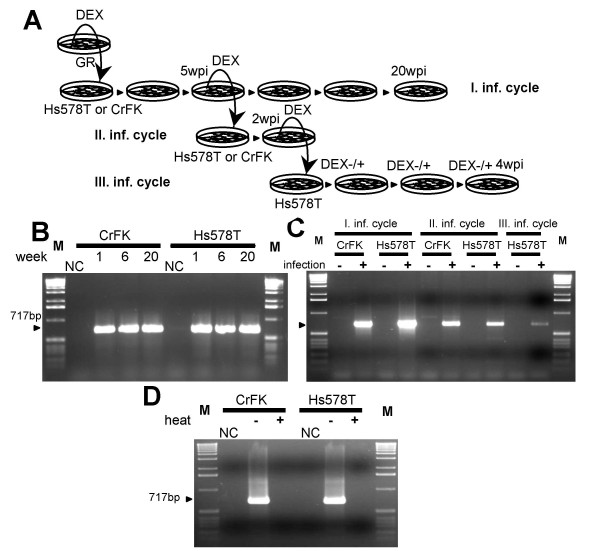
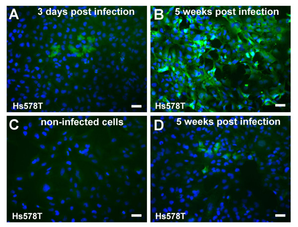
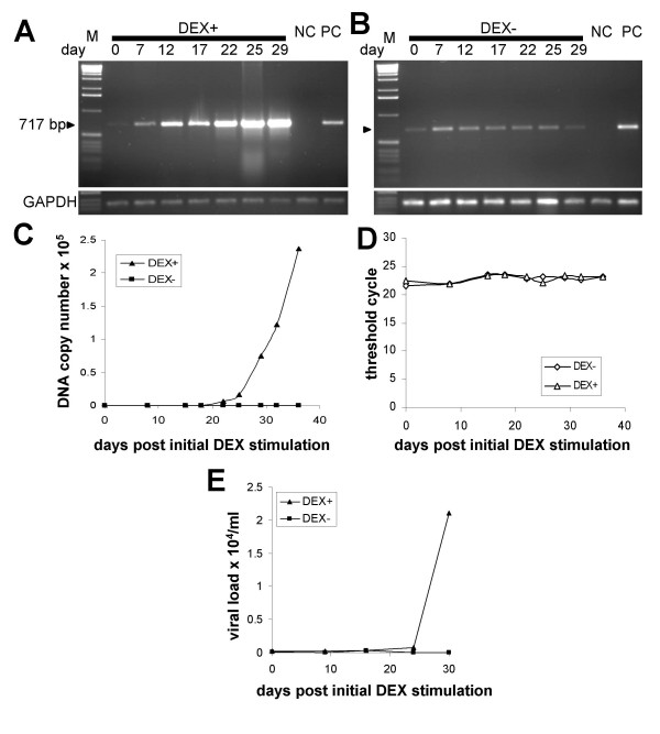
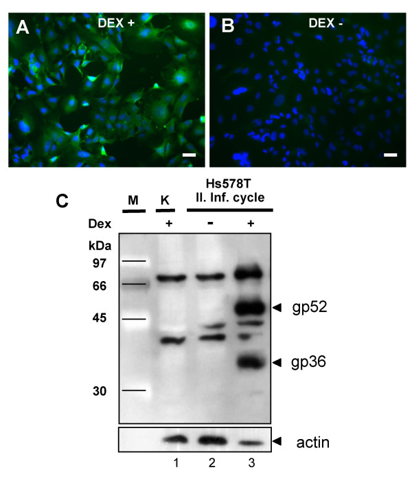
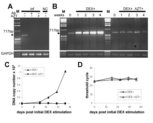
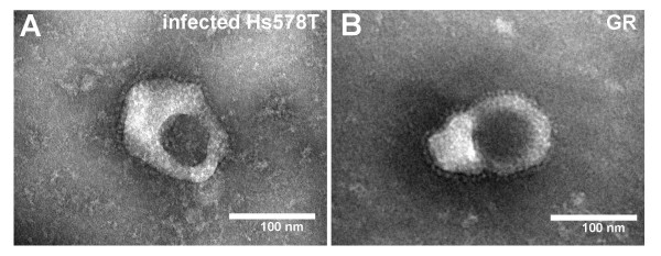
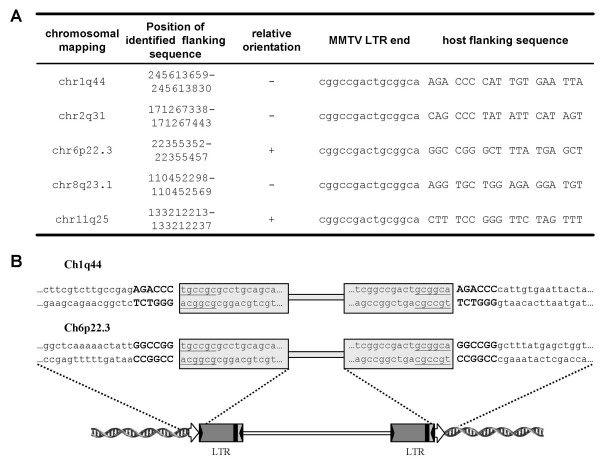
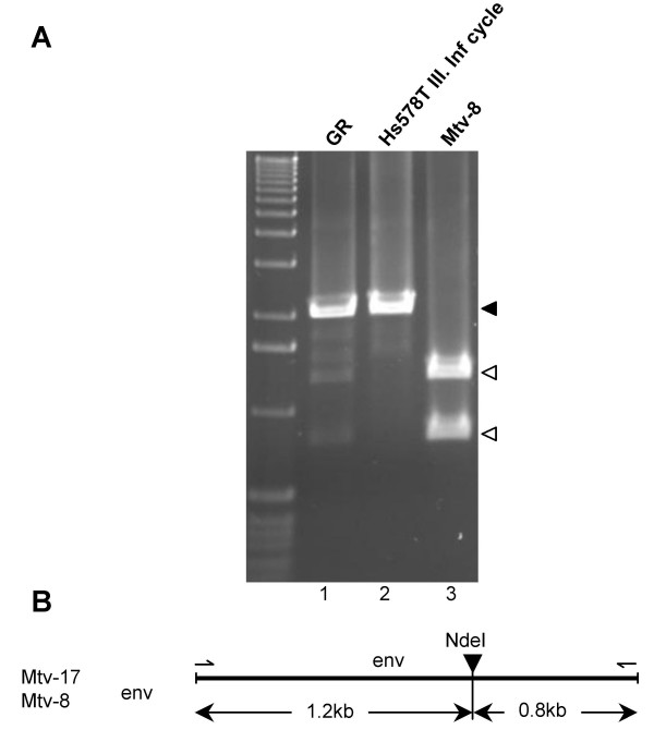
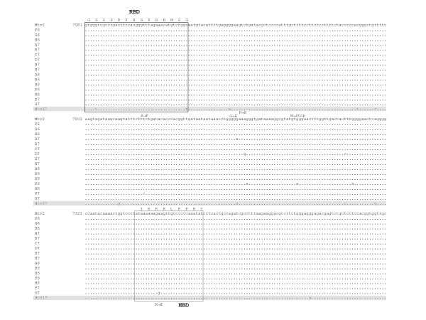
Results: Here, we show for the first time the rapid spread of mouse mammary tumor virus, MMTV(GR), in cultured human mammary cells (Hs578T), ultimately leading to the infection of every cell in culture. The replication of the virus was monitored by quantitative PCR, quantitative RT-PCR and immunofluorescence imaging. The infected human cells expressed, upon cultivation with dexamethasone, MMTV structural proteins and released spiked B-type virions, the infectivity of which could be neutralized by anti-MMTV antibody. Replication of the virus was efficiently blocked by an inhibitor of reverse transcription, 3'-azido-3'-deoxythymidine. The human origin of the infected cells was confirmed by determining a number of integration sites hosting the provirus, which were unequivocally identified as human sequences.

Conclusion: Taken together, our results show that human cells can support replication of mouse mammary tumor virus.


Figures

Figure 1
Figure 1
Infection of human breast cell line Hs578T and feline kidney cells, CrFK, with MMTV(GR) virus. (A) Experimental design. Wpi: weeks post infection. (B) The cells infected with MMTV(GR) were monitored for 20 weeks. Genomic DNA was harvested at week one, six and 20 after infection, respectively, and analyzed by PCR for the presence of MMTV sequences. NC: non-infected cells. M: 1 kb marker. (C) Three infectious cycles were performed in Hs578T cells. The cells infected with MMTV(GR) virus are denoted as first infection cycle. The cell culture supernatant from these Hs578T cells was used in a subsequent infection round. Medium from the second-cycle infected Hs578T cells was used for third infection cycle. M: 1 kb marker. (D) Heat inactivation of the MMTV(GR). Where indicated (heat +) was the virus subjected to the heat treatment (60°C for 10 min). NC: non-infected cells, M: 1 kb marker.
Figure 2
Figure 2
Immunofluorescence imaging of MMTV-infected Hs578T cells. The expression of capsid proteins in the third-round infected Hs578T cells was visualized by immunofluorescence staining using a monospecific anti-CA serum. Only a small number of MMTV-positive cells was detected in the third-round infected cells 3 days after infection (A), whereas by week five all the cells expressed MMTV antigen (B). The increase was strictly DEX dependent. Upon cultivation of the cells in DEX-free medium no increase in the number of CA-positive cells could be observed (D). (C) Non-infected Hs578T cells. 24 h prior immunostaining all the cells (A, B, C, D) were grown in medium supplemented with 10-6 M DEX. (Scale bar, 50 μm).
Figure 3
Figure 3
Quantification of proviral DNA and viral RNA in cell lysates and supernatants of the third-round infected human breast cells during a time-course experiment. (A and B) The third-round infected cells were cultured in the presence (A) or absence (B) of 10-6 M DEX. Genomic DNA was extracted from the infected cells at the indicated time points and semiquantitative PCR was performed. NC: non-transduced HS578T cells. PC: second-round infected Hs578T cells. Equal DNA loading was controlled in a PCR assay with GAPDH-specific primers (bottom panels). M: 1 kb marker. (C) Real-time TaqMan PCR quantifying proviral loads in the infected Hs578T cells during the time-course experiment. (D) Equal loading of the PCR reactions was controlled in a Real-time TaqMan PCR specific for GAPDH gene. (E) The viral RNA was quantified by Real-time RT-PCR in cell culture fluids of the infected Hs578T cells grown either in the presence or absence of 10-6 M DEX.
Figure 4
Figure 4
Detection of expression of MMTV proteins in the infected human cells. (A and B) Infected Hs578T cells were cultured for 5 weeks in the presence of 10-6 M DEX. One week before immunofluorescence staining the cells were cultivated in the absence of the glucocorticoid analog and 24 h prior immunofluorescence staining with anti-CA antibodies the production of MMTV-specific proteins was either induced (A) or not (B) by addition of 10-6 M DEX in the cell culture media. The nuclei of the cells were counterstained with DAPI. (C) Western blot detecting the expression of gp52 and gp36 Env proteins in the second-round infected HS578T cells. Lane 1, non-infected Hs578T cells, NC; lane 2, infected human cells not stimulated with DEX; lane 3, infected human cells in which the expression of MMTV structural proteins was induced by 10-6 M DEX 24 h before protein harvest.
Figure 5
Figure 5
Neutralization of viral infectivity and AZT treatment. (A) The presence of proviral DNA in the infected Hs578T cells was determined by PCR. The virus released from the second round infected Hs578T cells was, prior infection, pre-incubated either with anti-MMTV neutralizing antibody (Ab) or PBS. Where indicated AZT was added to the cells infected with the virus. NC: non-infected Hs578T cells. M: 1 kb marker. (B) Spread of the virus was abrogated in medium containing AZT. The third-round infected Hs578T cells were cultured for four weeks in medium containing DEX either supplemented with AZT or not and the presence proviral DNA was monitored by a semiquantitative PCR. GAPDH-specific PCR was used to demonstrate equal loading of all PCR reactions (bottom panels). M: 1 kb marker. (C) Real-time TaqMan PCR quantifying proviral loads in the infected Hs578T cells during the AZT treatment experiment. (D) Equal loading was contolled in a Real-time TaqMan PCR specific for GAPDH gene.
Figure 6
Figure 6
Electron microscopy of viral particles released from the infected Hs578T (A) and GR cells (B).
Figure 7
Figure 7
Virus-host junction sequences. (A) The junctions detected by LM-PCR in Hs578T cells infected with MMTV(GR). Terminal sequence of MMTV LTR (small letters) and 18 nucleotides of host flanking sequence (capital letters) are shown. Determined host sequence was mapped using a BLAT search at the UCSC Genome Bioinformatics group web page. The exact position of the host sequence amplified in LM-PCR on the chromosome is numbered according to Human Mar. 2006 (hg18) assembly. (B) Duplications of 6 bp long host provirus flanking sequences were determined. MMTV proviral sequences are boxed, inverted repeats at the end of LTRs are underlined. Duplications of host flanking sequences are indicated by large bold letters. Schematic diagram of an integrated MMTV provirus is shown below. Direct repeats of the host sequence are indicated by open arrows. Inverted repeats terminating the LTR are shown as inverted solid triangles.
Figure 8
Figure 8
Virus infecting human cells do not contain env gene derived from endogenous retroviruses. (A) The PCR products encompassing complete env coding region amplified from the cell lysates of GR cells (lane 1) and third-round infected Hs578T cells (lane 2) were submitted to digestion with NdeI. As a digestion control, PCR product obtained by amplification of MTV-8 env sequences using pGR16 plasmid as a template, was digested with the same restriction enzyme (lane 3); M, 1 kb marker; black arrow indicates undigested product; open arrows denote fragments resulting from NdeI digestion. (B) Schematic drawing showing NdeI site in the env gene of the Mtv-17 and Mtv-8 viruses and the length of the respective restriction fragments.
Figure 9
Figure 9
Alignment of a partial env gene sequences from the third infection cycle of Hs578T cells. Genomic DNA extracted from the third-round infected Hs578T cells, was used for amplification and cloning of MMTV env sequences. Sequences of sixteen clones were aligned. Mtv-2 and Mtv-17 (accession number AF263910, sequence is shaded) sequences were included in the alignment. Putative heparin binding domain (HBD) and receptor binding site (RBS) are boxed and amino acid residues representing these regions are shown above the boxes. Non-synonymous mutations in proviral sequences from infected cells resulting in an amino acid exchange are indicated. The coordinates of the Mtv2 nucleotide sequence are according to the MMTV reference strain (accession number M15122).

Similar articles

Cited by

References

    1. Sarkar NH. Type B virus and human breast cancer. In: Giraldo A and Beth A, editor. The role of viruses in human cancer. Vol. 1 , Elsevier, North Holland; 1980. pp. 207–235.
    1. Mant C, Cason J. A human murine mammary tumour virus-like agent is an unconvincing aetiological agent for human breast cancer. Rev Med Virol. 2004;14:169–177. doi: 10.1002/rmv.427. - DOI - PubMed
    1. Wang Y, Holland JF, Bleiweiss IJ, Melana S, Liu X, Pelisson I, Cantarella A, Stellrecht K, Mani S, Pogo BG. Detection of mammary tumor virus env gene-like sequences in human breast cancer. Cancer Res. 1995;55:5173–5179. - PubMed
    1. Etkind P, Du J, Khan A, Pillitteri J, Wiernik PH. Mouse mammary tumor virus-like ENV gene sequences in human breast tumors and in a lymphoma of a breast cancer patient. Clin Cancer Res. 2000;6:1273–1278. - PubMed
    1. Ford CE, Tran D, Deng Y, Ta VT, Rawlinson WD, Lawson JS. Mouse mammary tumor virus-like gene sequences in breast tumors of Australian and Vietnamese women. Clin Cancer Res. 2003;9:1118–1120. - PubMed

Publication types

MeSH terms