. 2007;35(20):6917-23.
doi: 10.1093/nar/gkm823. Epub 2007 Oct 11.

Extent to which hairpin opening by the Artemis:DNA-PKcs complex can contribute to junctional diversity in V(D)J recombination

Affiliations

Extent to which hairpin opening by the Artemis:DNA-PKcs complex can contribute to junctional diversity in V(D)J recombination

Haihui Lu et al. Nucleic Acids Res. 2007.

Abstract

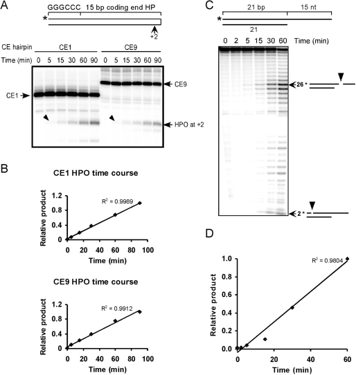
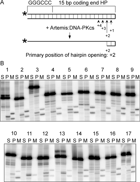
V(D)J recombination events are initiated by cleavage at gene segments by the RAG1:RAG2 complex, which results in hairpin formation at the coding ends. The hairpins are opened by the Artemis:DNA-PKcs complex, and then joined via the nonhomologous DNA end joining (NHEJ) process. Here we examine the opening of the hairpinned coding ends from all of the 39 functional human V(H) elements. We find that there is some sequence-dependent variation in the efficiency and even the position of hairpin opening by Artemis:DNA-PKcs. The hairpin opening efficiency varies over a 7-fold range. The hairpin opening position varies over the region from 1 to 4 nt 3' of the hairpin tip, leading to a 2-8 nt single-stranded 3' overhang at each coding end. This information provides greater clarity on the extent to which the hairpin opening position contributes to junctional diversification in V(D)J recombination.


Figures

Figure 1.
Figure 1.
The endonuclease activity of Artemis:DNA-PKcs is linear over a 90-min time course. (A) Two different coding end hairpins 1 and 9 (CE1 and CE9) were assayed for time courses of hairpin opening by Artemis:DNA-PKcs. The arrowheads point to the primary hairpin opening products at the +2 position. (B) The major products at each time point were quantified and normalized to the product at the last time point (90 min). A linear trend line was added and the R-squared value was displayed on the chart. (C) The time course of 3′ DNA overhang cleavage by Artemis:DNA-PKcs. The 3′ DNA overhang substrate was illustrated on the top. The major products are identified using arrows with the nt sizes and corresponding cleavage positions (arrowheads) depicted on the right. (D) The primary cleavage products of 26 nt at each time point were plotted in the same manner as in (B).
Figure 2.
Figure 2.
The phosphorylation of Artemis by DNA-PKcs rapidly reaches a plateau. (A) Artemis was incubated with DNA-PKcs in the presence of [γ-32P] ATP at 37°C. A fraction of the incubation mixture was removed at each time point and resolved on SDS–PAGE. For the zero time point, a separate reaction containing all components except DNA-PKcs was used. (B) The level of phosphorylation was quantified and normalized as a percentage of the highest level (100% at 30 min).
Figure 3.
Figure 3.
Hairpin opening by Artemis:DNA-PKcs as a function of KCl or Ku. (A) The substrate used here is coding end hairpin 1 (CE1). Proteins in the reactions are indicated above the corresponding lanes. Reactions 1–4 are done in 25 mM KCl, while reactions 5–8 are done in 75 mM KCl. To activate DNA-PKcs, reactions 1–7 included a 60 bp double-strand DNA with short overhangs, whereas reaction 8 instead contains a pseudo-Y structured DNA with a 20 bp double-strand region as depicted. The numbers along the bottom are the quantitation of hairpin opening (product/total). (B) Radioactive kinase assays with DNA-PKcs and Artemis were carried out at the indicated KCl concentrations, and Ku protein was added to reaction 5.
Figure 4.
Figure 4.
The hairpin opening pattern by Artemis:DNA-PKcs for the 17 different coding end hairpins derived from 39 human VH elements. (A) The illustration shows the configuration of coding end hairpin substrates and the cleavage positions by Artemis:DNA-PKcs. The base pairings are indicated as thin lines between the top and bottom strands. The length of the arrows reflects different cleavage efficiency at the four positions (+1 to +4). (B) The hairpin opening products were resolved by denaturing PAGE. The numbers 1–17 correspond to the sequence numbers in Table 1. For each coding end hairpin (HP), there is a set of three lanes. The hairpin opening products (P) were in the middle, with uncut substrate on the left (S), and the hairpin opening markers (M) on the right. For most substrates, the markers correspond to hairpin opening at +1 and +3 positions, except for substrate 11, with only one marker at the +2 position. CE3 substrate appears to show one minor hairpin opening product at greater than the +4 position.

References

    1. Swanson PC. The bounty of RAGs: recombination signal complexes and reaction outcomes. Immunol. Rev. 2004;200:90–114. - PubMed
    1. Fugmann SD, Lee AI, Shockett PE, Villey IJ, Schatz DG. The RAG proteins and V(D)J recombination: complexes, ends, and transposition. Ann. Rev. Immunol. 2000;18:495–527. - PubMed
    1. Tonegawa S. Somatic generation of antibody diversity. Nature. 1983;302:575–581. - PubMed
    1. Gellert M. V(D)J recombination: RAG proteins, repair factors, and regulation. Annu. Rev. Biochem. 2002;71:101–132. - PubMed
    1. Lieber MR. Site-specific recombination in the immune system. FASEB J. 1991;5:2934–2944. - PubMed

Publication types