. 2008 Feb;22(2):344-60.
doi: 10.1210/me.2007-0400. Epub 2007 Oct 11.

A repressive role for prohibitin in estrogen signaling

Affiliations

A repressive role for prohibitin in estrogen signaling

Bin He et al. Mol Endocrinol. 2008 Feb.

Abstract

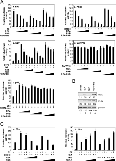
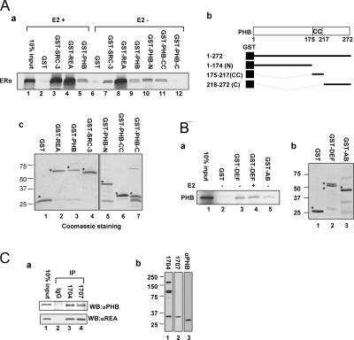
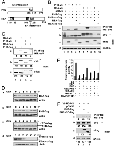
Nuclear receptor-mediated gene expression is regulated by corepressors and coactivators. In this study we demonstrate that prohibitin (PHB), a potential tumor suppressor, functions as a potent transcriptional corepressor for estrogen receptor alpha (ERalpha). Overexpression of PHB inhibits ERalpha transcriptional activity, whereas depletion of endogenous PHB increases the expression of ERalpha target genes in MCF-7 breast cancer cells. Chromatin immunoprecipitation experiments demonstrate that PHB is associated with the estrogen-regulated pS2 promoter in the absence of hormone and dissociates after estradiol treatment. We demonstrate that PHB interacts with the repressor of estrogen receptor activity (REA), a protein related to PHB, to form heteromers and enhance the protein stability of both corepressors. Interestingly, the corepressor activity of PHB is cross-squelched by the coexpression of REA (and vice versa), suggesting that PHB and REA repress transcription only when they are not paired. We further demonstrate that coiled-coil domains located in the middle of PHB and REA are responsible for their heteromerization, stabilization, and cross-squelching actions. Finally, ablation of PHB function in the mouse results in early embryonic lethality, whereas mice heterozygous for the PHB null allele exhibit a hyperproliferative mammary gland phenotype. Our results indicate that PHB functions as a transcriptional corepressor for ERalpha in vitro and in vivo, and that its heteromerization with REA acts as a novel mechanism to limit its corepressor activity.


Figures

Figure 1
Figure 1
Prohibitin Represses ERα-Mediated Transcription A, PHB and REA are corepressors for ERα, PR-B, and E2F1, but not for Gal4-VP16 and p53. Transcriptional activities of ERα, PR-B, E2F1, Gal-VP16, and p53 were determined by cotransfection of HepG2 cells with increasing amounts of REA, PHB, or REA/PHB (50, 100, 200, and 300 ng). B, Western blot analysis to show the expression of PHB and REA protein from their respective vectors. The 293T cells were transiently transfected with empty vector (lane 1), or vector expressing REA (lane 2) or PHB (lane 3), or both expression vectors (lane 4). Expression levels were determined by using anti-REA, anti-PHB antibodies. β-Actin was analyzed as sample loading control. C, PHB and REA antagonize ERα coactivation by SRC-3. In the left panel, 300 ng of SRC-3 was cotransfected with increasing amounts of REA or PHB plasmids (100, 200, and 300 ng) in HepG2 cells. On the other hand, overexpression of SRC-3 overcomes the ERα activity repressed by PHB and REA. In right panel, 200 ng of REA or PHB were cotransfected with increasing amounts of SRC-3 (100, 200, and 300 ng).
Figure 2
Figure 2
ERα, PHB and REA Directly Interact with Each Other A-a, GST pull-down shows a direct interaction between ERα and PHB, which is hormone independent. The interaction domains of PHB with ERα map to the NH2 terminus and the CC domain. As a control, the interaction between SRC-3 and ERα is enhanced by E2 treatment. A-b, Schematic diagram of PHB, indicating the location of the NH2 terminus [amino acids (aa) 1–174, N], CC domain (aa 175–217, CC), and carboxyl terminus (aa 218–272, C) is shown. A-c, Amounts of each GST fusion protein used in the pull-down assays. Stars indicate the expected sizes for each expressed proteins. B-a, Reciprocal GST pull-down experiment confirms a direct interaction between ERα and PHB in a hormone-independent manner. B-b, The amounts of each GST fusion protein used. AB, NH2-terminal regulatory domain that contains aa 1–180; DEF, includes hinge region, ligand binding domain, and C-terminal variable region, which contains aa 251–595. C-a, Protein extract from MCF7 cells were immunoprecipitated with two antibodies specifically against different epitopes of REA. Western blot analysis demonstrated the in vivo association of PHB with REA. As a negative control, nonspecific IgG could not precipitate PHB. C-b, In Western blot (WB) analysis, anti-REA antibodies BL1704 and BL1707 do not recognize PHB, whereas an anti-PHB antibody (rabbit polyclonal; Santa Cruz Biotechnology) can only recognize PHB.
Figure 3
Figure 3
Depletion of Endogenous PHB Increases the Expression of ERα Target Genes in MCF-7 Cells A-a and A-b, siRNA against PHB and REA specifically depleted the mRNA of PHB and REA in MCF-7 cells, respectively, as determined by real-time PCR. A-c, The protein levels of PHB and REA were simultaneously reduced by siRNA against either PHB or REA, indicating that PHB and REA depend on each other for protein stability. B, In MCF-7 cells, expression of ERα target gene pS2 was strongly increased by depletion of REA, and moderately increased by depletion of PHB (B-a). Expression of cyclin D1 mRNA was significantly increased by depletion of PHB, but not by depletion of REA (B-b). Individual siRNA against REA mRNA nos. 2 and 3 caused a strong increase of pS2 gene expression, whereas siRNA nos. 1 and 4 only show slight effect (B-c). This is in agreement with the knock down efficiency of REA protein level with individual siRNAs (B-e). Similarly individual siRNAs against PHB mRNA nos. 2–4 caused a moderate but statistically significant increase of cyclin D1 (CCND1) gene expression, whereas siRNA no. 1 only show little effect (B-d). Shown here is a representative of three experiments (±sd). *, P < 0.05). This again is in agreement with the knock down efficiency of PHB protein level by these individual siRNAs (B-f). C, Recruitment of PHB to endogenous pS2 gene promoter. MCF-7 cells were grown in stripped media for 3 d, and treated with vehicle or 10 nm E2 for 45 min. ChIP assays were conducted with antibodies against ERα, SRC-3, and PHB. The input lanes represent for 5% of total genomic DNA used in ChIP assay. RNAi, RNA interference.
Figure 4
Figure 4
The CC Domains of PHB and REA Are Required for Hetero-Oligomerization, Stabilization, and Transcriptional Cross-Squelching A, Schematic diagram of functional domains of PHB and REA. B, Transient expression of carboxyl-terminal V5 or flag tagged PHB and REA wild-type and CC deletion mutants in 293T cells. Protein extracts were first immunoprecipitated (IP) with anti-flag antibody, and precipitated protein complexes were analyzed by Western blot (WB) analysis with an anti-V5 antibody. Bands in lanes 1–5 in B-a represent the immunoprecipitated V5-tagged PHB, whereas bands in lanes 6–10 represent the immunoprecipitated V5-tagged REA. B-b through -d, Input control of different tagged PHB and REA wild type and mutants and β-actin. C, REA-V5, PHBV5, REA-flag, and PHB-flag were overexpressed in 293T cells separately. D, Flag-tagged PHB and REA wild types or CC deletion mutants were expressed in 293T cells. The protein synthesis inhibitor cycloheximide (CHX) was added to a final concentration of 200 μg/ml and cells were harvested at the indicated time points. Protein stability was measured by Western blot analysis with anti-flag antibody (top panels of D-a through -d), and anti-β-actin antibody as an input control (bottom panels of D-a through -d). E, The CC domains are required for the cross-squelching actions between PHB and REA. The HepG2 cells were cotransfected with ERE-luc reporter vector, expression vectors of ERα (5 ng), REAΔCC (300 ng), PHBΔCC (300 ng), REAΔ/PHBΔ (150 ng of REAΔCC and 150 ng of PHBΔCC), REA (100 ng), PHB (100 ng), REA/PHB (50 ng of REA and 50 ng of PHB), REA (300 ng), PHB (300 ng), or REA/PHB (150 ng of REA and 150 ng of PHB). F, V5-HDAC1, PHB-flag, and PHBΔCC were overexpressed in 293T cells separately. Western blot analysis was used to determine the amounts of cell lysates that contain equal levels of PHB wild type and PHBΔCC. After this, the cell lysates containing either contain PHB-flag or PHBΔCC-flag was mixed with equal amounts of cell lysates containing V5-HDAC1. After overnight incubation, the cell lysates were immunoprecipitated with beads linked to anti-flag antibodies, and probed with antibodies against V5.
Figure 5
Figure 5
Generation of PHB Knockout Mice A, PHB gene trapped ES cell clone XT0035 was obtained from Sanger Institute Gene Trap Resource. The gene-trap targeting vector is comprised of the En2 intron, a splice acceptor site, a β-geo fusion cassette composed of β-galactosidase and the neomycin resistance marker, and a simian virus 40 polyadenylation signal. The targeting vector was inserted into the coding region of last exon. B, The ES cells were stained with X-gal. The blue staining indicates PHB gene promoter is active in ES cells. C, Southern blot analysis using neo probe to demonstrate the single locus insertion of the target vector. The genomic DNA purified from the ES clone XT0035 were digested with NsiI (lane 1) and SpeI (lane 2). Neither the restriction enzyme NsiI nor SpeI cuts the gene trap vector. The predicted sizes of restriction fragments resulting from NsiI and SpeI digestion are 14.6 and 16.7 kb, respectively. D, Epithelial cells were isolated from inguinal mammary glands of wild-type (WT) and PHB+/− mice, and total proteins were extracted, subject to Western blot (WB) analysis with the antibodies against PHB, REA, and β-actin. Each lane represents the protein extract from the epithelial cells isolated from two mice. E, The wild-type and PHB+/− female siblings were treated with a standard 3-wk E-P treatment regimen, which induces mammary gland ductal side branching and alveologenesis. Proteins were extracted from the whole mammary glands. Western blot analysis was conducted using different antibodies against cyclin D1 (CCND1), PHB, REA, and β-tubulin (E-a). The relative protein levels of REA, PHB, and cyclin D1 were quantitated and shown in E-b.
Figure 6
Figure 6
H&E and Immunohistochemistry Staining of E6.5 Decidual Balls E6.5 decidual balls isolated from PHB+/− females mated with PHB+/− male were fixed in 10% formaldehyde, sectioned, and subject to H&E and immunohistochemistry staining. A-a and A-c, The H&E staining of a representative decidual ball containing a normally developed E6.5 embryo at two different magnifications. A-b and A-d, H&E staining of a representative decidual ball which does not contain noticeable embryo. PDZ, SDZ, and EM denote the primary decidual zone, secondary decidual zone, and embryo, respectively. B-a and B-c, Immunohistochemistry staining of a representative E6.5 embryo with anti-PHB antibody. B-b and B-d, Negative control staining in which the anti-PHB primary antibody was not included. B-e, Anti-PHB staining of another section of E6.5 embryo. B-f, Anti-β-galactosidase staining of the consecutive section of E6.5 embryo used for B-e. exec, Extraembryonic ectoderm; ep, epiblast; pac, pro-amniotic cavity.
Figure 7
Figure 7
The Mammary Glands of PHB Heterozygous Mice Showed Hyper-Proliferation after E-P Treatment A and B, Whole mounts of inguinal mammary glands from E-P-treated wild type (WT) (A) and PHB+/− (B) mice, respectively (LN, lymph node). C and D, Higher magnifications of regions of panels A and B, respectively. E and F, H&E-stained sections of mammary glands shown in panels A and B, respectively. Compared with the E-P-treated wild-type mouse gland, note the significant increase in alveologenesis and ductal side branching (black arrowhead) in the E-P-treated PHB+/− mouse gland. G, Alveolar bud number per field (±sd). **, P < 0.01) in E-P-treated wild-type and PHB+/− mouse glands. H and I, 5-Bromo-2-deoxyuridine (BrdU) incorporation in mammary gland sections. H, Luminal epithelial cells of wild-type mammary gland, whereas panel I shows the luminal epithelial cells of PHB+/− mammary gland. J, Average percentages of mammary epithelial cells scoring positive for BrdU staining (±sd). *, P < 0.05) in E-P-treated wild-type and PHB+/− mouse glands.
Figure 8
Figure 8
Working Model for Multifunctional Proteins PHB and REA PHB and REA are able to interact with each other to form hetero-oligomers. There is equilibrium between homomer and heteromer forms of PHB and REA. We propose that, only when they are not paired, PHB and REA can function as transcriptional regulators for a variety of transcriptional factors, including ERα (30), E2F1 (36), PR-B, and AR (59). On the other hand, the hetero-oligomers of PHB and REA exert other functions, such as mitochondrial chaperones (40) and B-cell receptor-associated proteins (71). Moreover, it has been reported that PHB acts as a vascular marker of adipose tissue (72), inhibitor of pyruvate carboxylase (73), propigmentation effector (74), and plays critical role in Ras signaling pathway (75). It remains unknown whether REA is also involved in these biological processes as PHB partner.

Similar articles

Cited by

References

    1. Evans RM 1988 The steroid and thyroid hormone receptor superfamily. Science 240:889–895 - PMC - PubMed
    1. Tsai MJ, O’Malley BW 1994 Molecular mechanisms of action of steroid/thyroid receptor superfamily members. Annu Rev Biochem 63:451–486 - PubMed
    1. Glass CK, Rosenfeld MG 2000 The coregulator exchange in transcriptional functions of nuclear receptors. Genes Dev 14:121–141 - PubMed
    1. Lonard DM, O’Malley BW 2006 The expanding cosmos of nuclear receptor coactivators. Cell 125:411–414 - PubMed
    1. O’Malley BW 2006 Molecular biology. Little molecules with big goals. Science 313:1749–1750 - PubMed

Publication types

MeSH terms