. 2008 Feb;22(2):263-72.
doi: 10.1210/me.2007-0324. Epub 2007 Oct 11.

CBP Is a dosage-dependent regulator of nuclear factor-kappaB suppression by the estrogen receptor

Affiliations

CBP Is a dosage-dependent regulator of nuclear factor-kappaB suppression by the estrogen receptor

Kendall W Nettles et al. Mol Endocrinol. 2008 Feb.

Abstract

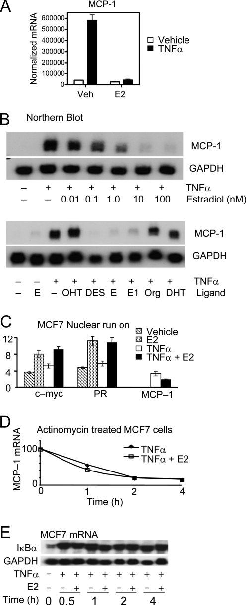
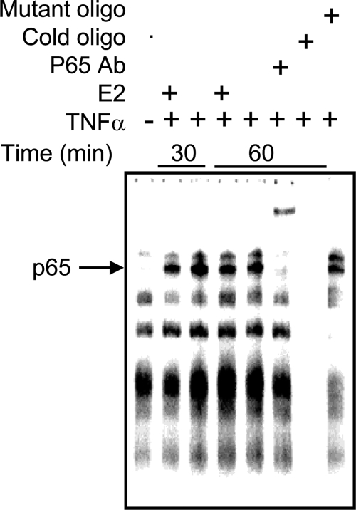
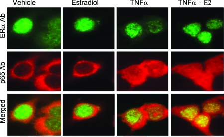
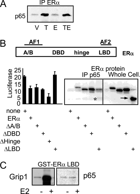
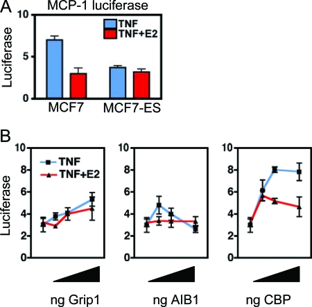
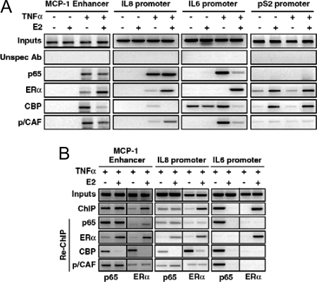
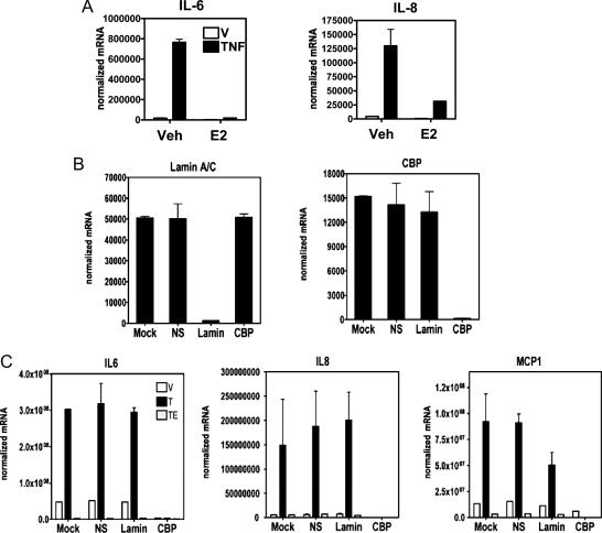
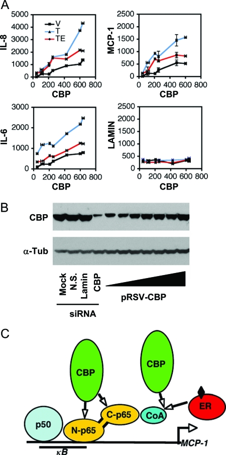
The estrogen receptor (ER) protects against debilitating effects of the inflammatory response by inhibiting the proinflammatory transcription factor nuclear factor-kappaB (NFkappaB). Heretofore cAMP response element-binding protein (CREB)-binding protein (CBP) has been suggested to mediate inhibitory cross talk by functioning either as a scaffold that links ER and NFkappaB or as a required cofactor that competitively binds to one or the other transcriptional factor. However, here we demonstrate that ER is recruited to the NFkappaB response element of the MCP-1 (monocyte chemoattractant protein-1) and IL-8 promoters and displaces CBP, but not p65, in the MCF-7 breast cancer cell line. In contrast, ER displaced p65 and associated coregulators from the IL-6 promoter, demonstrating a gene-specific role for CBP in integrating inflammatory and steroid signaling. Further, RNA interference and overexpression studies demonstrated that CBP dosage regulates estrogen-mediated suppression of MCP-1 and IL-8, but not IL-6, gene expression. This work further demonstrates that CBP dosage is a critical regulator of gene-specific signal integration between the ER- and NFkappaB-signaling pathways.


Figures

Figure 1
Figure 1
ERα Suppresses NFκB-Dependent MCP-1 Transcription A, MCF-7 cells were treated with TNFα and/or E2 for 2 h and then processed for qPCR analysis. The mRNA levels were normalized to 18S mRNA. B, Northern blot of MCP-1 and GAPDH mRNA expression. MCF-7 cells were cultured for 3 d in steroid-depleted media and stimulated for 2 h with 50 ng/ml TNFα (as indicated). Cultures were supplemented with steroid receptor ligands at 10 nm unless otherwise specified: E, estradiol; OHT, 4-hydroxytamoxifen; 100 nm 4-hydroxytamoxifen; DES, diethylstilbestrol; E1, estrone; ORG 2058, a synthetic progestin; DHT, dihydrotestosterone. These blots are representative of four independent experiments. C, Nuclei from MCF-7 cells were isolated and subject to run-on transcription assays to measure levels of transcription. D, Actinomycin was used to arrest transcription in MCF-7 cells. MCP-1 mRNA was normalized to GADPH mRNA. E, Northern blot of IκBα and GAPDH mRNA from MCF-7 cells treated with the indicated ligands. PR, progesterone receptor; Veh, vehicle.
Figure 2
Figure 2
Gel Shift of Radiolabeled NFκB Response Element Oligonucleotide from MCP-1 Gene MCF-7 Cells Were Treated for 30 or 60 min and Then Made into Nuclear Extracts, Demonstrating that E2 Has No Effect on DNA Binding by p65. Ab, Antibody.
Figure 3
Figure 3
Immunofluorescence Analysis of ERα and p65 MCF-7 cells were grown on coverslips, treated for 30 min with ligands, methanol fixed, and stained for immunofluorescent microscopy. The TNFα treatment induced a diffuse nuclear and cytoplasmic staining for p65, as previously reported for MCF-7 cells (31). Ab, Antibody.
Figure 4
Figure 4
Cellular Associations between ERα and p65 A, Immunoprecipitation of MCF-7 cell extracts, treated with ligands for 30 min before cell lysis. A polyclonal ERα antibody (ER21) was used for precipitation, followed by Western blotting for p65 (Santa Cruz anti-p65 A20). B (left panel), Cos-1 cells were transfected with MCP-1 luciferase reporter and ERα expression plasmids. The next day, cells were treated with TNFα + E2 for 6 h and processed for luciferase activity; B (right panel), Cos-1 cells were transfected with p65 and ERα expression plasmids as indicated. After 48 h, cells were treated for 30 min with TNFα + E2 and lysed for protein extraction. Only the LBD deletion construct was unable to interact with p65 (asterisk), although it was expressed at high levels in the whole-cell extract (arrow). C, A GST-ERα LBD fusion protein bound to glutathione-Sepharose beads was treated with vehicle or 1 mm E2 for 1 h and then used to pull down in vitro-translated Grip1 or p65. After extensive washing, the bound proteins were eluted with glutathione and visualized with SDS-PAGE and autoradiography. AF2, Activation function 2; IP, immunoprecipitation; DBD, DNA-binding domain; V, vehicle; T, TNFα; E, estradiol.
Figure 5
Figure 5
Role of CBP in ERα-NFκB Cross Talk A, An MCP-1-promoter-luciferase reporter demonstrates the repressive effects of E2 on the MCP-1 gene. Cells were transfected with the reporter and treated with Vehicle, TNFα, or TNFα + E2. Shown is the fold induction of the MCP-1 promoter activity relative to vehicle. We identified a clonal variant of MCF-7 cells that do not show the suppressive effects of E2, termed MCF-7-ES cells. B, MCP-1 promoter activity was measured in MCF-7-ES cells, which showed no suppression by E2 in the absence of added coactivator. The luciferase activity is shown relative to vehicle control. This luciferase reporter was cotransfected with increasing amounts of coactivator expression vector, or pBluescript to normalize the total DNA transfected. The next day, cells were treated for 6 h with the indicated ligands. Each data point was performed in triplicate, showing mean + sem, and the experiments were repeated four to five times. AIB1, Amplified in breast cancer 1.
Figure 6
Figure 6
CBP Displacement Mediates Estrogen-Dependent Gene Repression A, Confluent MCF-7 cells were switched to media with charcoal-stripped serum for 3 d. Cells were then treated with TNFα and E2, as indicated for 3 h before formaldehyde fixation, and subject to ChIP analysis. Engagement of the depicted proteins onto pS2 control promoter and MCP-1 enhancer was evaluated by using cognate antibodies and relative quantitative PCR. B, Re-ChIP analysis evaluating the simultaneous presence of the indicated proteins within complexes engaged onto the MCP-1 enhancer region. Unspec Ab, Unspecified antibody.
Figure 7
Figure 7
RNA Interference Targeting CBP A, RT-qPCR analysis of gene expression. MCF-7 cells were treated for 2 h with the indicated ligands and processed for analysis of the indicated gene expression, which was normalized to expression of 18S mRNA. B, RT-qPCR analysis of CBP or lamin gene expression. MCF-7 cells were transfected for 40 h with the indicated siRNA and then processed for qPCR analysis of lamin A/C or CBP, which is shown normalized to 18S mRNA. C, Effects of siRNA targeting CBP on inflammatory gene expression. MCF-7 cells were transfected with the indicated siRNAs for 40 h and then treated with vehicle, TNFα, or TNFα+E2 for 2 h, and then processed for qPCR analysis of gene expression. NS, Normal serum; TE, Tris-EDTA; Veh, vehicle; V, vehicle; T, TNFα.
Figure 8
Figure 8
CBP Is a Dosage-Sensitive Regulator of ER-NFκB Cross Talk A, MCF-7 cells were transfected with siRNA targeting CBP and varying amounts of pRSV-CBP expression vector. The next day, cells were treated for 2 h with vehicle, TNFα (15 ng/ml), and/or E2 (100 nm), and then processed for qPCR analysis of gene expression. After normalizing all mRNA levels to 18S, the target gene expression was expressed relative to the mRNA levels of CBP. B, Model of CBP interactions with the NFκB enhancer in the MCP-1 gene. CBP has well-characterized interactions with the N-terminal domain of CBP. ER does not interact directly with p65 and thus may compete with CBP binding through some other component of the assembled complex (see text for details). CoA, Coenzyme A; N.S., normal serum; α-Tub, α-tubulin.

References

    1. Koehler KF, Helguero LA, Haldosen LA, Warner M, Gustafsson JA 2005 Reflections on the discovery and significance of estrogen receptor β. Endocr Rev 26:465–478 - PubMed
    1. Chadwick CC, Chippari S, Matelan E, Borges-Marcucci L, Eckert AM, Keith Jr JC, Albert LM, Leathurby Y, Harris HA, Bhat RA, Ashwell M, Trybulski E, Winneker RC, Adelman SJ, Steffan RJ, Harnish DC 2005 Identification of pathway-selective estrogen receptor ligands that inhibit NF-κB transcriptional activity. Proc Natl Acad Sci USA 102:2543–2548 - PMC - PubMed
    1. Jimi E, Ghosh S 2005 Role of nuclear factor-κB in the immune system and bone. Immunol Rev 208:80–87 - PubMed
    1. Ghosh S, Karin M 2002 Missing pieces in the NF-κB puzzle. Cell 109 (Suppl):S81–S96 - PubMed
    1. Hayden MS, Ghosh S 2004 Signaling to NF-κB. Genes Dev 18:2195–2224 - PubMed

Publication types

MeSH terms