. 2008 Feb;22(2):234-47.
doi: 10.1210/me.2007-0124. Epub 2007 Oct 11.

Human immunodeficiency virus (HIV)-1 viral protein R suppresses transcriptional activity of peroxisome proliferator-activated receptor {gamma} and inhibits adipocyte differentiation: implications for HIV-associated lipodystrophy

Affiliations

Human immunodeficiency virus (HIV)-1 viral protein R suppresses transcriptional activity of peroxisome proliferator-activated receptor {gamma} and inhibits adipocyte differentiation: implications for HIV-associated lipodystrophy

Shashi Shrivastav et al. Mol Endocrinol. 2008 Feb.

Abstract

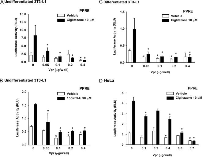
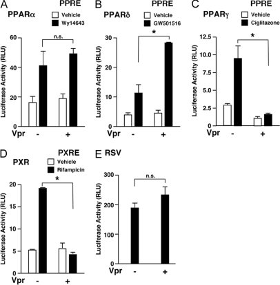
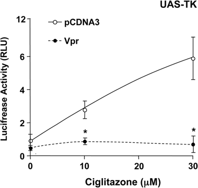
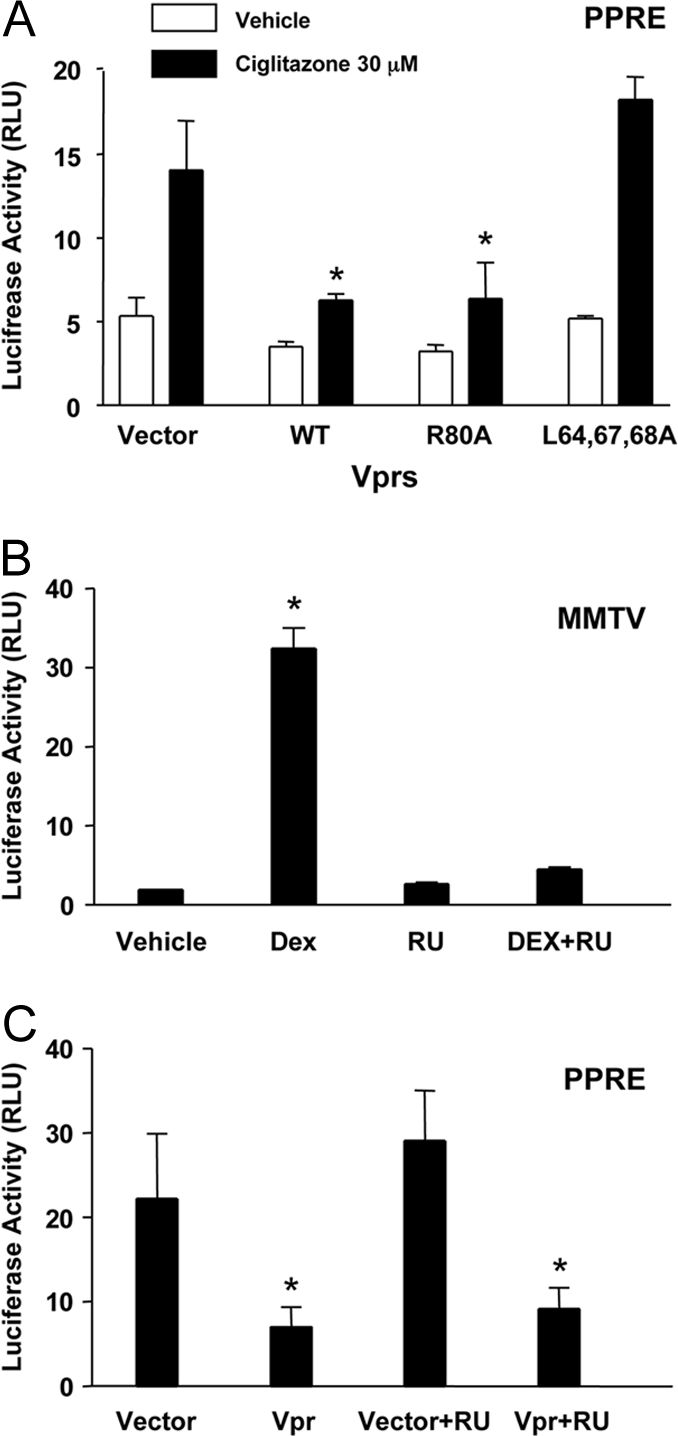
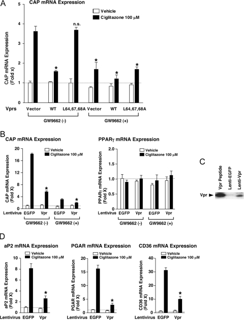
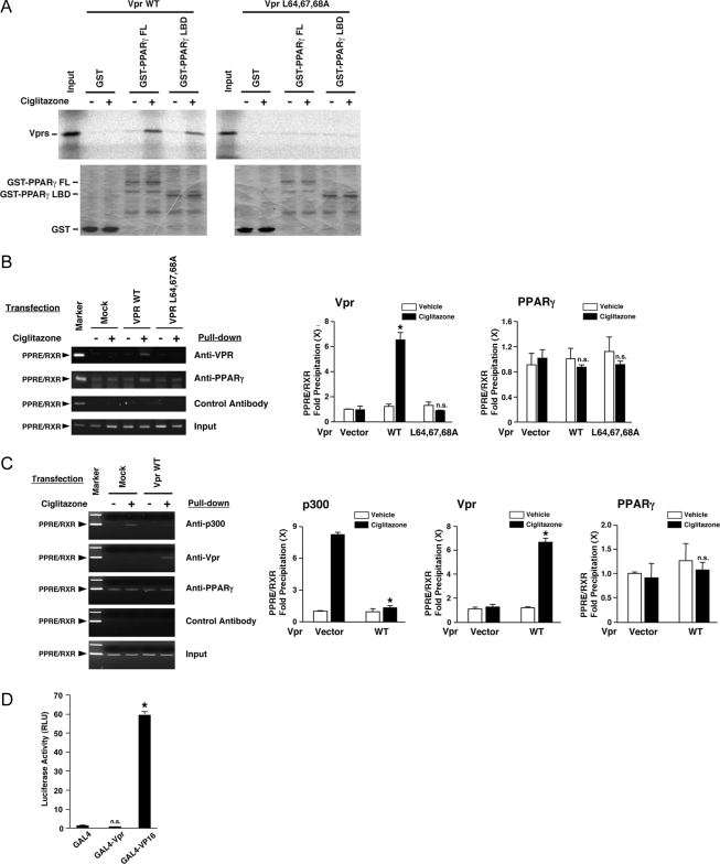
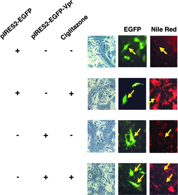
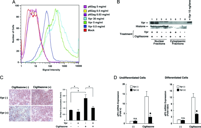
HIV-1-infected patients may develop lipodystrophy and insulin resistance. We investigated the effect of the HIV-1 accessory protein viral protein R (Vpr) on the activity of the peroxisome proliferator-activating receptor-gamma (PPARgamma), a key regulator of adipocyte differentiation and tissue insulin sensitivity. We studied expression of PPARgamma-responsive reporter genes in 3T3-L1 mouse adipocytes. We investigated Vpr interaction with the PPAR/retinoid X receptor (RXR)-binding site of the c-Cbl-associating protein (CAP) gene using the chromatin immunoprecipitation assay as well as the interaction of Vpr and PPARgamma using coimmunoprecipitation. Finally, we studied the ability of exogenous Vpr protein to enter cultured adipocytes and retard differentiation. We found that Vpr suppressed PPARgamma-induced transactivation in both undifferentiated and differentiated 3T3-L1 cells. Transcriptional suppression by Vpr required an intact LXXLL coactivator motif. Vpr suppressed mRNA expression of PPARgamma-responsive genes in undifferentiated 3T3-L1 cells and associated with the PPAR/RXR-binding site located in the promoter region of the CAP gene. Vpr interacted with the ligand-binding domain of PPARgamma in an agonist-dependent fashion in vitro. Vpr delivered either by an expression plasmid or as protein added to media suppressed PPARgamma agonist-induced adipocyte differentiation, assessed as lipid accumulation and mRNA expression of the adipocyte differentiation marker adipocyte P2 in 3T3-L1 cells. In conclusion, circulating Vpr or, alternatively, Vpr produced as a consequence of direct infection of adipocytes could suppress in vivo differentiation of preadipocytes by acting as a corepressor of PPARgamma-mediated gene transcription. Vpr may alter sensitivity to insulin and thereby contribute to the development of lipodystrophy and insulin resistance observed in HIV-1-infected patients.


Figures

Figure 1
Figure 1
Vpr Suppressed PPARγ-Induced Transcription in Undifferentiated (A and B) and Differentiated (C) 3T3-L1 and HeLa (D) Cells Cells were transfected with Vpr, PPARγ, and RXRγ expression plasmids, together with the reporter gene PPRE-TK-Luc and the normalizing gene pCMV-β-Gal.Bars represent mean ± sem values of the luciferase activity normalized for β-galactosidase activity in the absence or presence of ciglitazone (A, C, and D) or 15d-PGJ2 (B). Vpr suppressed PPRE-mediated gene expression in a dose-dependent fashion in the presence of the PPARγ ligands ciglitazone and 15-dPDJ, in both murine adipocytes (3T3-L1 cells) and human cervical carcinoma HeLa cells. *, P < 0.01 compared with baseline (0 in the presence of ciglitazone).
Figure 2
Figure 2
Vpr Has Different Transcriptional Effects on PPARγ, PPARα, PPARδ, and PXR-Mediated Transcription in Undifferentiated 3T3-L1 Cells Cells were transfected with Vpr- and RXRγ-expressing plasmids, PPRE-TK-Luc, and pCMV-β-Gal together with PPARα-expressing (A), PPARδ-expressing (B) or PXR-expressing (C) plasmids. The Vpr-expressing plasmid RSV-Luc and pCMV-β-Gal were transfected into the cells (D). Bars represent mean ± sem values of the luciferase activity normalized for β-galactosidase activity in the absence or presence of Wy14643 (A), GW501516 (B), ciglitazone (C), or rifampicin (D). *, P < 0.01; n.s., not significant.
Figure 3
Figure 3
Vpr Suppressed PPARγ-Induced Transactivation in a GAL4-DBD-Mediated Mammalian One-Hybrid System in Undifferentiated 3T3-L1 Cells Cells were transfected with a plasmid that expresses PPARγ fused to a GAL4-DBD, together with a response plasmid pUAS-TK-Luc and the normalizing plasmid pCMV-β-Gal. Data represent mean ± sem of luciferase activity normalized for β-galactosidase activity. There was a dose-dependent increase in gene expression, which suggests that Vpr interacts with PPARγ, and this interaction is facilitated by the PPARγ ligand ciglitazone. *, P < 0.01 comparing the presence and absence of Vpr.
Figure 4
Figure 4
Vpr Suppressed PPARγ-Induced Transactivation via Its LXXLL Motif Independently of Its Enhancement of GR Transcriptional Activity A, Vpr suppressed PPARγ-induced transactivation through an LXXLL motif. Undifferentiated 3T3-L1 cells were cotransfected with indicated amounts of PPARγ, RXRγ, PPRE-TK-Luc, and pCMV-β-Gal and with mutant and Vpr-expression plasmids. Bars represent values of the luciferase activity normalized for β-galactosidase activity in the absence or presence of ciglitazone. Mutant Vpr R80A, deficient in cell cycle-arrest activity, was able to suppress PPARγ-induced transactivation in undifferentiated 3T3-L1 cells. In contrast, triple-mutant Vpr lacking the corepressor motif was unable to suppress PPARγ-induced transactivation. *, P < 0.01, compared with baseline (vector in the presence of ciglitazone). B and C, Vpr suppressed PPARγ-induced transactivation independently of its coactivation of glucocorticoid-mediated gene transcription. Cells were cotransfected with PPARγ, RXRγ-pCMV-β-Gal, and MMTV-Luc expression plasmids (B) or PPRE-TK-Luc (C) in the presence or absence of Vpr-expression plasmid. Cells were subsequently treated with dexamethasone and/or RU 486 (B) and ciglitazone (C). Bars represent mean ± sem. Values of the luciferase activity were normalized for β-galactosidase. *, P < 0.01; n.s., not significant compared with the baseline (vector).
Figure 5
Figure 5
Vpr Suppressed mRNA Expression of the Endogenous PPARγ-Responsive CAP Gene in Undifferentiated 3T3-L1 Cells A, Wild-type Vpr, but not the L64,67,68A mutant, suppressed the CAP mRNA expression in undifferentiated 3T3-L1 cells. Cells were transfected with Vpr and PPARγ- and RXRγ-expression plasmids and treated with ciglitazone and/or the PPARγ antagonist GW9662 for 24 h. Total RNA was purified from the cells, and the expression of CAP and GAPDH mRNAs was determined by real-time PCR. Bars show mean ± sem of the fold induction of CAP normalized for GAPDH, compared with baseline (vector in the presence of ciglitazone and in the absence of GW9662). Ciglitazone stimulation of CAP expression was blunted by wild-type Vpr and in the presence of GW9662 but not by triple-mutant Vpr lacking the corepressor motif. B, Vpr expressed by lentivirus infection strongly suppressed ciglitazone-induced CAP mRNA expression in undifferentiated 3T3-L1 cells. Cells were infected with the lentiviruses that express Vpr or EGFP and were treated with ciglitazone and/or GW9662 for 24 h. Total RNA was purified from the cells, and the expression of CAP, PPARγ, and GAPDH mRNAs was determined by real-time PCR. Bars show mean ± sem of the fold induction of CAP or PPARγ normalized for GAPDH, compared with baseline (EGFP in the presence of ciglitazone and in the absence of GW9662). C, Vpr was expressed in cells infected with the lentivirus vector for Vpr. Cells were infected with lentiviruses expressing Vpr or EGFP, and cell lysates were run on a 4–20% SDS-PAGE gel. Vpr peptide (10 ng) was run as a control. After blotting on a nitrocellulose membrane, Vpr was visualized with the anti-Vpr antibody. D, Vpr expressed by lentivirus infection suppressed the expression of the ciglitazone-induced PPARγ-responsive aP2, PAGP, and CD36 mRNAs in undifferentiated 3T3-L1 cells. The same samples used in Fig. 5B were examined. aP2, PGAR, CD36, and GAPDH mRNAs were determined by real-time PCR. Bars show mean ± sem of the fold induction of aP2, PGAR, or CD36 mRNAs normalized for GAPDH, compared with baseline (EGFP) in the presence of ciglitazone.
Figure 6
Figure 6
Vpr Interacted with PPARγ in Vitro and in Vivo Vpr attenuates attraction of p300 coactivator to the PPARγ-responsive promoter, whereas it does not have autonomous transactivation or transrepression activity in undifferentiated 3T3-L1 cells. A, Wild-type Vpr, but not LXXLL defective Vpr mutant, bound the LBD of PPARγ in a ligand-dependent fashion in a GST pull-down assay. In vitro translated and labeled wild-type Vpr or VprL64,67,68A was incubated with bacterially produced GST-fused full-length PPARγ or PPARγ LBD immobilized on GST beads. Results of the GST pull-down assay are shown in the top panels, whereas expression of the GST-fusion proteins is shown in the bottom panels. B, Wild-type Vpr, but not L64,67,68A mutant, was attracted to the PPRE/RXR-binding site of the CAP promoter in a ciglitazone-dependent fashion in undifferentiated 3T3-L1 cells. Cells were transfected with wild-type Vpr, VprL64,67,68A, and PPARγ- and/or RXRγ-expression plasmids and treated with ciglitazone or vehicle. Twenty-four hours after addition of ciglitazone, the cells were fixed, and the ChIP reaction was performed with anti-Vpr, anti-PPARγ, or control antibody. The portion of the CAP promoter that contains one PPAR/RXR-binding site was amplified by PCR. Results obtained in the SYBR-Green real-time PCR for quantitatively evaluating the ChIP results are shown in the right panels. Bars represent mean ± se values of fold precipitation of the CAP PPRE/RXR-binding site, compared with the baseline. *, P < 0.01; n.s., not significant, compared results to baseline (vector in the presence of ciglitazone). C, Wild-type Vpr attenuated ciglitazone-induced association of p300 to the CAP promoter in undifferentiated 3T3-L1 cells. Cells were transfected with Vpr and PPARγ- and RXRγ-expression plasmids and treated with ciglitazone or vehicle. Twenty-four hours after addition of ciglitazone, the cells were fixed, and the ChIP reaction was performed with anti-Vpr, anti-PPARγ, anti-p300, or control antibody. The portion of the CAP promoter that contains one PPAR/RXR-binding site was amplified by PCR with a specific primer pair, and obtained gel images on 3% DNA gels are shown in the left panel. Results obtained in the SYBR-Green real-time PCR for quantitatively evaluating the ChIP results are shown in the right panels. Bars represent mean ± se values of fold precipitation of the CAP PPRE/RXR-binding site, compared with the baseline. *, P < 0.01; n.s., not significant, compared with the baseline (vector in the presence of ciglitazone). D, Vpr does not have intrinsic transactivation or transrepression activity in undifferentiated 3T3-L1 cells. Undifferentiated 3T3-L1 cells were transfected with pM, pM-Vpr, or pGLA4-Vpr16, together with GAL4-responsive p17mer-tk-Luc and CMV-β-galactosidase. Bars show the means ± sem. *, P < 0.01; n.s., not significant, compared with the baseline (GAL4).
Figure 7
Figure 7
Vpr Delivered by Transfection Suppresses Ciglitazone-Induced Lipid Accumulation and Differentiation of 3T3-L1 Cells Undifferentiated 3T3-L1 cells were transfected with pIRES2-EGFP-Vpr or the control vector pIRES2-EGFP and treated with 10 μm ciglitazone or vehicle for 48 h as indicated. Nomarski image and fluorescence signals from EGFP and lipid stained with Nile red were obtained. Ciglitazone enhanced cellular lipid uptake, whereas Vpr-expressing cells failed to accumulate lipid in response to ciglitazone. Arrows in the two panels are directed at cells that have taken up the Vpr-expressing plasmid (presence of green fluorescence) but are lacking the differentiated adipocyte phenotype (absence of lipid accumulation).
Figure 8
Figure 8
Extracellularly Administered Vpr Suppressed PPARγ-Induced Lipid Accumulation in 3T3-L1 Cells A, Undifferentiated 3T3-L1 cells took up extracellularly administered fluorescence-labeled Vpr peptide in a dose-dependent fashion. 3T3-L1 cells were incubated with indicated concentrations of fluorescein isothiocyanate-labeled Vpr or p6Gag and cells with the fluorescein isothiocyanate-labeled Vpr were sorted by fluorescence-activated cell sorting. B, Western blot analysis detected Vpr in the nucleus of undifferentiated 3T3-L1 cells exposed to the synthetic Vpr peptide. Undifferentiated 3T3-L1 cells were incubated with 100 ng/ml Vpr and/or 10 μm ciglitazone for 5 d, and nuclear and cytoplasmic fractions were analyzed by Western blot using antibodies against Vpr and histone 1 (the latter to confirm nuclear fractionation). C, Extracellularly administered Vpr inhibits ciglitazone-induced lipid accumulation in 3T3-L1 cells. 3T3-L1 cells were cultured with 100 ng/ml synthetic Vpr peptide and/or 10 μm ciglitazone for 5 d. The cells were analyzed for lipid accumulation with Oil Red O staining. The left panel indicates representative images of Oil Red O staining, whereas the right panel demonstrated accumulation of Oil Red O-stained area. Bars show mean ± sem of the fold induction of the Oil Red O-stained area. *, P < 0.05. Magnification, ×10. D, Extracellularly administered synthetic Vpr peptide suppresses ciglitazone-induced aP2 mRNA expression in undifferentiated (left panel) and differentiated (right panel) 3T3-L1 cells. Undifferentiated and differentiated 3T3-L1 cells were incubated with 100 ng/ml synthetic Vpr peptide and/or 10 μm ciglitazone for 5 d. Total RNA was purified from the cells, and mRNA expression of aP2 and RPLP0 were determined by real-time PCR. Cell differentiation by ciglitazone resulted in a higher concentration of aP2 mRNA as compared with undifferentiated cells treated with ciglitazone. Bars show mean ± sem of the fold induction of aP2 mRNA expression normalized for those of RPLP0. *, P < 0.01; n.s., not significant, by comparison between results obtained in the absence and presence of ciglitazone.

Similar articles

Cited by

References

    1. Carr A, Samaras K, Burton S, Law M, Freund J, Chisholm DJ, Cooper DA 1998 A syndrome of peripheral lipodystrophy, hyperlipidaemia and insulin resistance in patients receiving HIV protease inhibitors. AIDS 12:F51–F58 - PubMed
    1. Sweet DE 2005 Metabolic complications of antiretroviral therapy. Top HIV Med 13:70–74 - PubMed
    1. Lichtenstein KA 2005 Redefining lipodystrophy syndrome: risks and impact on clinical decision making. J Acquir Immune Defic Syndr 39:395–400 - PubMed
    1. Rudich A, Ben-Romano R, Etzion S, Bashan N 2005 Cellular mechanisms of insulin resistance, lipodystrophy and atherosclerosis induced by HIV protease inhibitors. Acta Physiol Scand 183:75–88 - PubMed
    1. Boufassa F, Dulioust A, Lascaux AS, Meyer L, Boue F, Delfraissy JF, Sobel A, Goujard C 2001 Lipodystrophy in 685 HIV-1-treated patients: influence of antiretroviral treatment and immunovirological response. HIV Clin Trials 2:339–345 - PubMed

Publication types

MeSH terms