. 2008 Jan 15;17(2):225-39.
doi: 10.1093/hmg/ddm300. Epub 2007 Oct 11.

Mutation in the splicing factor Hprp3p linked to retinitis pigmentosa impairs interactions within the U4/U6 snRNP complex

Affiliations

Mutation in the splicing factor Hprp3p linked to retinitis pigmentosa impairs interactions within the U4/U6 snRNP complex

Juana Maria Gonzalez-Santos et al. Hum Mol Genet. .

Abstract

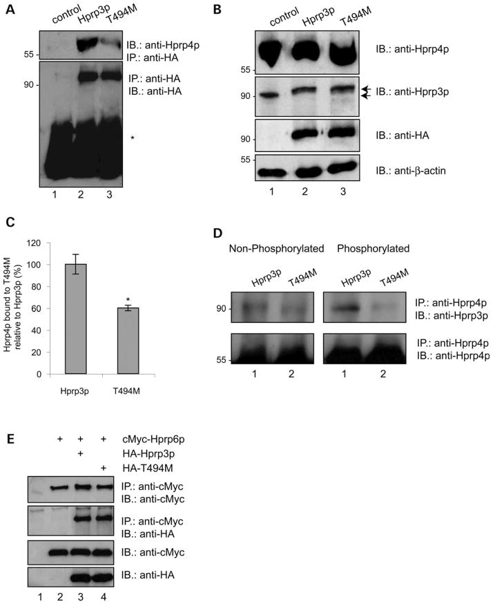
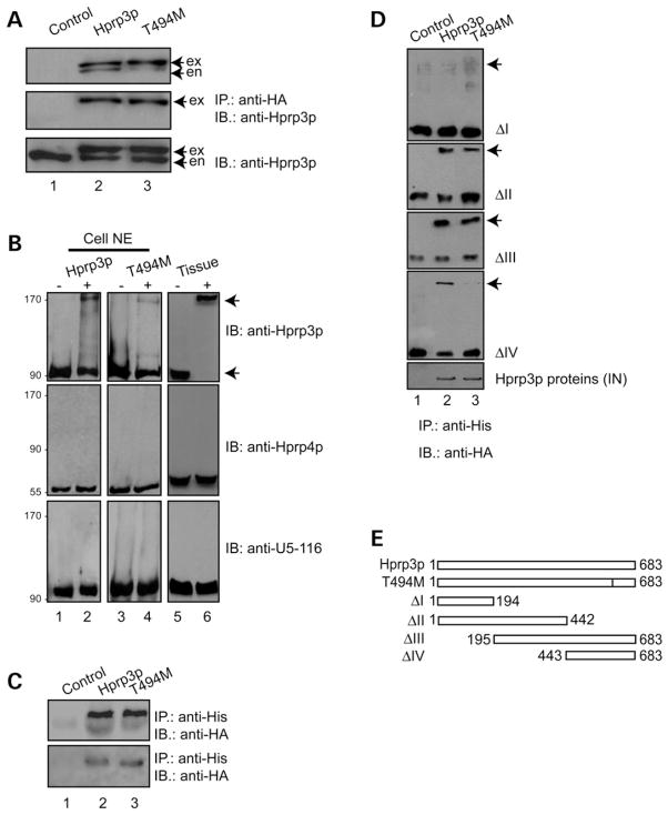
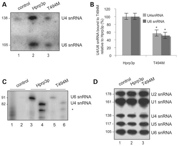
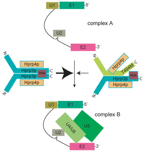
Mutations in PRPF3, a gene encoding the essential pre-mRNA splicing factor Hprp3p, have been identified in patients with autosomal dominant retinitis pigmentosa type 18 (RP18). Patients with RP18 have one of two single amino acid substitutions, Pro493Ser or Thr494Met, at the highly conserved Hprp3p C-terminal region. Pro493Ser occurs sporadically, whereas Thr494Met is observed in several unlinked RP families worldwide. The latter mutation also alters a potential recognition motif for phosphorylation by casein kinase II (CKII). To understand the molecular basis of RP18, we examined the consequences of Thr494Met mutation on Hprp3p molecular interactions with components of the U4/U6.U5 small nuclear ribonucleoprotein particles (snRNPs) complex. Since numerous mutations causing human diseases change pre-mRNA splice sites, we investigated whether Thr494Met substitution affects the processing of PRPF3 mRNA. We found that Thr494Met does not affect PRPF3 mRNA processing, indicating that the mutation may exert its effect primarily at the protein level. We used small hairpin RNAs to specifically silence the endogenous PRPF3 while simultaneously expressing HA-tagged Thr494Met. We demonstrated that the C- but not N-terminal region of Hprp3p is indeed phosphorylated by CKII in vitro and in cells. CKII-mediated Hprp3p phosphorylation was significantly reduced by Thr494Met mutation. Consequently, the Hprp3p C-terminal region is rendered partially defective in its association with itself, Hprp4p, and U4/U6 snRNA. Our findings provide new insights into the biology of Hprp3p and suggest that the loss of Hprp3p phosphorylation at Thr494 is a key step for initiating Thr494Met aberrant interactions within U4/U6 snRNP complex and that these are likely linked to the RP18 phenotype.


Conflict of interest statement

Conflict of Interest statement. None declared.

Figures

Figure 1
Figure 1
Putative high-score SR protein motifs in exon 11 of PRPF3 are abrogated by C1482T mutation. (A) ESE motif scores for exon 11 of PRPF3 wild-type (left) and T494M (right) were calculated by ESEfinder (5,6,9,53). High-score motifs are in dark gray for SF2/ASF, and light gray for SRp40. The height of each bar indicates the score value, the position along the x axis indicates its location along the exon 11 and the width of the bar represents the length of the motif. The C at position 1482 in PRPF3 is underlined. The T at the same position in T494M causes both SF2/ASF and SRp40 scores to fall below threshold (2.87 to −0.075 and 3.26–1.54, respectively). Thresholds and maximal values are different for different SR proteins (1.95 for SF2/ASF and 2.67 for SRp40). (B) Schematic of the minigenes used in this study. PRPF3 and T494M minigenes were cloned into pcDNA3, using EcoRI (E) and XhoI (X) restriction endonucleases. Horizontal arrows under pcDNA3 and exon 12 indicate forward and reverse primers for PCR, respectively. (C) Analysis of PRPF3 and T494M minigenes. RT–PCR was carried out to assess pre-mRNA splicing from PRPF3 (lane 2), and T494M (lane 3) minigene constructs or empty vector (lane 1). The RT–PCR products were resolved by 1.5% agarose. Histone 1(0) gene served as an internal control. DNA size loading marker is shown on the left.
Figure 2
Figure 2
Expression of wild-type and mutant Hprp3p in cells when the endogenously expressed Hprp3p is knocked down. (A) Schematic of the complementation system designed to silence the endogenous PRPF3 gene while expressing its wild-type or mutant versions in cells. An HD-Ad vector was designed to produce two shRNAs, which silence expression of the endogenous PRPF3 by targeting both the 5′- and 3′-UTRs of its mRNA. The vector also expresses either the exogenous wild-type PRPF3: HD-Ad-F3iplus (upper) or T494M: HD-Ad-F3iT494M (lower), both lacking the targeted sequences. (B) Expression of T494M 4 days post-transduction with HD-Ad viral vector. Western blots were performed with antibodies against Hprp3p (upper) or HA (middle). β-actin was used as loading control (lower). exo, exogenous HA-Hprp3p; en, endogenous Hprp3p. Numbers on the left refer to size in kDa. (C) Indirect immunostaining micrographs show expression and subcellullar localization of the HA-Hprp3p protein in cells 4 day post-transduction. Vectors used for transduction are indicated on the right: empty vector (HD-Ad; a and b), HD-Ad-F3i which expresses shRNAs only (c and d), HD-Ad-F3iplus (e and f), and HD-Ad-F3iT494M (g and h). Identical fields are shown for DAPI (left) and FITC (right) channels. The nuclear DNA is visualized as blue fluorescence (a, c, e and g). The localization of the HA-tagged protein is visualized as green immunofluorescence (f and h). Mock-transfected cells did not display any immunostaining signal under the FITC channel (b and d). Fluorescence micrographs were recorded using a 100× objective.
Figure 3
Figure 3
CKII-mediated Hprp3p phosphorylation is weakened by substitution of Thr494 for Met. (A) Diagram of Hprp3p, T494M and truncated constructs (mutants I, and IV) shows potential threonine phosphorylation sites as predicted by NetPhos and Scansite programs. (B) CKII mediates Hprp3p phosphorylation in cells. Nuclear extracts, transduced with HA-tagged Hprp3p or T494M, were subjected to immunoprecipitation using antibody against HA in the absence (lanes 1, 2 and 4) or presence (lanes 3 and 5) of heparin. As negative control, nuclear extract from non-transduced cells was used (lane 1). Threonine phosphorylation was detected by western blotting with antibodies against phosphothreonine (upper). For loading control, membranes were stripped and reprobed with anti-HA antibody (lower). Results are representative of at least three experiments. *IgG heavy chains. (C and D) CKII phosphorylates Hprp3p in vitro. His-tagged Hprp3p, T494M (C) or Hprp3p truncated versions—ΔI, ΔIII and ΔIII-T494M (D) were incubated with CKII in the presence of GTP. Representative bar graph (C, lower) shows the decrease levels on threonine phosphorylation of T494M mutant protein relative to Hprp3p (*P < 0.01). The proteins were separated on 8–10% SDS–PAGE and visualized by western blotting with antibodies specific to phosphothreonine antibody (upper). Membranes were stripped and reprobed with anti-HA antibody (lower). *background bands. anti-HA, anti-HA antibody; anti-P-thr, anti-phosphothreonine antibody.
Figure 4
Figure 4
Effect of T494M mutation on Hprp3p binding to Hprp4p and Hprp6p. (A) Hprp3p/Hprp4p interactions is weakened by substitution of Thr494 for Met. Nuclear extracts were prepared from ARPE-19 cells 4 days after transduction with HD-Ad viral vectors that silence endogenous Hprp3p and express intact HA-Hprp3p (upper, lane 2), HA-T494M (lane 3). The extracts were immunoprecipitated using anti-HA monoclonal antibody bound to protein G-agarose beads. 20% of immunoprecipitates were resolved by 8% SDS–PAGE and immunoblotted using anti-Hprp4p polyclonal antibody. Control (lane 1) represents cells transduced with empty HD-Ad vector. The blot shown in A (upper) was stripped and reprobed with anti-HA antibody (lower). *IgG heavy chains. (B) The level of total nuclear proteins was analyzed by direct immunoblotting. 5% of nuclear extract input, used for immunoprecipitation in A, was resolved as above and probed with anti-Hprp4p (upper), Hprp3p (second, arrows indicate size difference between endogenous Hprp3p and HA-Hprp3p), anti-HA antibodies (third) or anti-β-actin as a loading control (lower). (C) Percentage of Hprp4 bound to T494M relative to Hprp3p was calculated by NIH Image software (v 1.62). (D) Phosphorylation of Hprp3p increases its binding to Hprp4p. E. coli expressed Hprp3p and T494M were phosphorylated in vitro using recombinant CKII and GTP (see Fig. 3C). After phosphorylation, the proteins were mixed with Hprp4p and co-IP using anti-Hprp4p antibody. In parallel, unphosphorylated His-tagged proteins were co-immunoprecipitated in a similar way. The immunoprecipitates were resolved by 8% SDS–PAGE and immunoblotted using anti-HA antibodies (top). The blots were stripped and reprobed with anti-Hprp4p antibody (bottom). (E) Hprp3p/Hprp6p association is not affected by T494M mutation. co-IP of HA-tagged Hprp3p and T494M by cMyc-Hprp6p. Cells co-expressing cMyc-Hprp6p and HA-Hprp3p or HA-T494M were co-IP with anti-cMyc antibody and the immunocomplexes fractionated by 8% SDS–PAGE (top). Nuclear extracts from cells transfected with empty vectors or expressing Hprp6p alone were used as controls (lanes 1 and 2, respectively).
Figure 5
Figure 5
Hprp3p self-association is affected by T494M substitution. (A) Hprp3p self-association is mediated by RNA in cells. NE from ARPE-19 cells transduced with HD-Ad-F3iplus, HD-Ad-F3iT494M or empty vector were immunoprecipitated with anti-HA antibody and the precipitants analyzed by western blotting with anti-Hprp3p antibody (top). Similar immunoprecipitation and immunoblotting analysis were performed using NE pretreated with RNase A (middle). The levels of each protein in the cell extracts are shown (lower). (B) Cross-linker induces Hprp3p self-association in cells and in animal tissue. DFDNB cross-linking of nuclear extracts from cells expressing Hprp3p (top left), T494M (top center) or animal tissue (top right). Middle and lower panels represent the same blots as above after probing with anti-Hprp4p or anti-U5-116 antibody as controls. (C) Hprp3p self-association is not mediated by Hprp4p. His-tagged, E. coli translated Hprp3p pulled-down either HA-Hprp3p or T494M from ARPE-19 NE in which Hprp4p was immunoblocked (top) or from NE pretreated with RNase A (lower). (D) T494M mutant protein does not associate with Hprp3p C-terminal region. Co-IP using His-tagged, E. coli translated truncated Hprp3p versions (mutants I, II, III and IV) and NE from ARPE-19 cells expressing either HA-Hprp3p or HA-T494M (arrows on the right). Input and precipitated proteins were resolved by 8 or 10% SDS–PAGE and probed with antibody against Hprp3p. NE, nuclear extracts; co-IP, co-immunoprecipitation; IN, input control showing 10% of the amount of the nuclear extract used in the co-IP. (E) Schematic shows Hprp3p, T494M and truncated Hprp3p versions (mutants I, II, III and IV).
Figure 6
Figure 6
U4 and U6 snRNAs association with Hprp3p is weakened by T494M mutation. (A and B) Binding of T494M mutant protein to U4/U6 snRNA. From the remaining 80% of immunoprecipitates mentioned in Fig. 4A, total RNA was extracted and the presence of U4 and U6 snRNAs detected by northern blot (A) and primer extension analysis (B). The migration position of U4 (138 or 82 nucleotides) and U6 (105 or 91nucleotides) are indicated on the right. *partial extension product from U4 snRNA. (C) Percentage of U4/U6 snRNAs bound to T494M relative to Hprp3p as detected by northern blot was calculated by NIH Image software (v 1.62). (D) Levels of U1, U2, U4, U5 and U6 snRNAs in 5 μg of total RNA isolated from ARPE-19 nuclear extracts used for immunoprecipitation are shown. SnRNAs were separated on 8% polyacrylamide-urea (8 M) gel, hybridized to oligonucleotide probes complementary to U1, U2, U4, U5 and U6 snRNAs and hybridization signals detected using a phosphoImager. RNAs were visualized by phosphoImager, analyzed using NIH Image software (v 1.62) and normalized to HA-Hprp3 proteins (for immunocomplexes) or the internal U1 control (for RNA inputs).
Figure 7
Figure 7
Effect of T494M missense mutation on cell proliferation, viability and global pre-mRNA splicing. Cell proliferation and viability were assessed by the MTT (A), trypan blue exclusion (B) and LDH cytotoxicity assay (C), after 2, 4 or 6 days post-transduction with HD-Ad-LacZ, HD-Ad-Fi, HD-Ad-F3iplus or HD-Ad-F3iT494M. Each experiment (mean ± SEM) was carried out in triplicate (n = 4). Statistical significance was assessed by one-way ANOVA and Holm’s multiple comparison test (*P < 0.05). The result is expressed in MTT activity (A), cell viability (B) and LDH release (C) relative to the corresponding treatment at day 0. (D) Expression of T494M 6 day post-transduction with HD-Ad viral vector. Western blots were performed with antibodies against Hprp3p (upper), HA (middle) or β-actin (bottom). exo, exogenous HA-Hprp3p; en, endogenous Hprp3p. (E) Indirect immunostaining micrographs show expression and subcellular localization of the HA-Hprp3p protein in ARPE-19 cells 6 day post-transduction with HD-Ad-F3iplus (see Fig. 2C, for labels). (F) Expression of RPE-specific and house-keeping genes in cells expressing wild-type or T494M mutant protein. RT–PCR was performed with total RNA isolated from ARPE-19 cells 6 day post-transduction. RPL18, EEF1A1, RPE65 and H1(0) mRNAs transcripts are shown. The DNA size marker is shown on the right.
Figure 8
Figure 8
Thr494 modulates Hprp3p association with the U4/U6-snRNP complex during the assembly of the B complex. The schematic shows a representation of the spliceosome assembly cycle, indicating the proposed role for Hprp3p in promoting the addition of the U4/U6 snRNP to the A complex through its association with other components of the spliceosome (thick arrow on the left). Substitution of Thr494 for Met may destabilize U4/U6 snRNP assembly by weakening Hprp3p association with U4/U6 snRNP components (thin arrow on the right). RNA, U4/U6 snRNA; U1, U2 and U5 indicate the corresponding snRNP; E, exon; A, intron branch site.

Similar articles

Cited by

References

    1. Wang Q, Chen Q, Zhao K, Wang L, Traboulsi EI. Update on the molecular genetics of retinitis pigmentosa. Ophthalmic Genet. 2001;22:133–154. - PubMed
    1. Daiger SP, Bowne SJ, Sullivan LS. Perspective on genes and mutations causing retinitis pigmentosa. Arch Ophthalmol. 2007;125:151–158. - PMC - PubMed
    1. Faustino NA, Cooper TA. Pre-mRNA splicing and human disease. Genes Dev. 2003;17:419–437. - PubMed
    1. Hamel C. Retinitis pigmentosa. Orphanet J Rare Dis. 2006;1:40. - PMC - PubMed
    1. McKie AB, McHale JC, Keen TJ, Tarttelin EE, Goliath R, van Lith-Verhoeven JJ, Greenberg J, Ramesar RS, Hoyng CB, Cremers FP, et al. Mutations in the pre-mRNA splicing factor gene PRPC8 in autosomal dominant retinitis pigmentosa (RP13) Hum Mol Genet. 2001;10:1555–1562. - PubMed

Publication types

MeSH terms

Grants and funding