. 2007 Nov;8(11):1052-60.
doi: 10.1038/sj.embor.7401088. Epub 2007 Oct 12.

Proteomic and functional analysis of Argonaute-containing mRNA-protein complexes in human cells

Affiliations

Proteomic and functional analysis of Argonaute-containing mRNA-protein complexes in human cells

Julia Höck et al. EMBO Rep. 2007 Nov.

Abstract

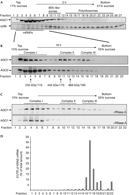
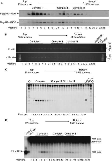
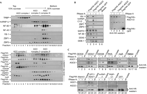
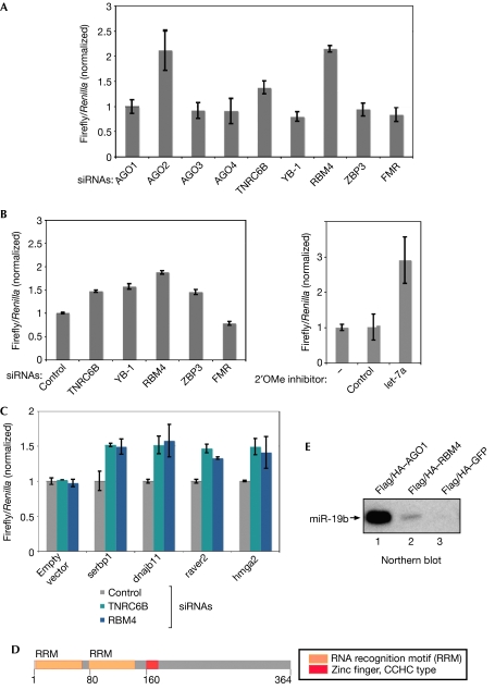
Members of the Argonaute (Ago) protein family associate with small RNAs and have important roles in RNA silencing. Here, we analysed Ago1- and Ago2-containing protein complexes in human cells. Separation of Ago-associated messenger ribonucleoproteins (mRNPs) showed that Ago1 and Ago2 reside in three complexes with distinct Dicer and RNA-induced silencing complex activities. A comprehensive proteomic analysis of Ago-containing mRNPs identified a large number of proteins involved in RNA metabolism. By using co-immunoprecipitation experiments followed by RNase treatment, we biochemically mapped interactions within Ago mRNPs. Using reporter assays and knockdown experiments, we showed that the putative RNA-binding protein RBM4 is required for microRNA-guided gene regulation.


Figures

Figure 1
Figure 1
Human AGO1 and AGO2 associate with distinct protein–RNA complexes. (A) Individual fractions of polyribosome gradients were analysed by western blotting against endogenous AGO1 (upper panel) or rpS6 (lower panel). (B) Lysates from wild-type HEK 293 cells were separated by sucrose density centrifugation under conditions that allow the separation of mRNPs. Endogenous AGO1 and AGO2 were analysed using specific antibodies. (C) Lysates were analysed as in (B). Lysates shown in the lower panel were treated with 100 μg/ml RNase A before centrifugation. (D) A reporter construct containing the KRAS 3′-UTR was transfected into HEK 293 cells and lysates were separated as in (B). Total RNA was extracted from the individual fractions and analysed by qRT–PCR. The distribution of the KRAS 3′-UTR is shown as a percentage of the total amount of the KRAS 3′-UTR. AGO, Argonaute; HEK, human embryonic kidney; KRAS, Kirsten rat sarcoma viral oncogene homologue; mRNPs, messenger ribonucleoproteins; qRT–PCR, quantitative reverse transcription–PCR; rpS6, ribosomal protein S6; 3′-UTR, 3′-untranslated region.
Figure 2
Figure 2
Argonaute complexes associate with distinct Dicer and RISC activities. (A) HEK 293 cell extracts containing Flag/HA–AGO1 (upper panel) or Flag/HA–AGO2 (lower panel) were separated by gradient centrifugation. The presence of Flag/HA–AGO1 and Flag/HA–AGO2 was analysed by western blotting using HA antibodies. (B) Lysates from HEK 293 cells expressing Flag/HA–AGO1 or Flag/HA–AGO2 were separated as in (A). Fractions were immunoprecipitated using Flag antibodies. RNA was extracted and the presence of endogenous let-7a (upper panel) and miR-16 (lower panel) was determined using RT–PCR. (C) Lysates from Flag/HA–AGO2-transfected HEK 293 cells were separated and immunoprecipitated as described in (A). Immunoprecipitates were incubated with a 32P-cap-labelled RNA, which contained a perfect complementary sequence to the endogenous miR-19b. Lanes indicated with T1 show RNase T1 digestions of the RNA substrates. The RNA sequence complementary to miR-19b is indicated by a black bar to the right. (D) Flag/HA–AGO1-containing HEK 293 lysate was separated and immunoprecipitated as described in (A). The immunoprecipitates or recombinant Dicer were incubated with an internally labelled pre-miR-27a substrate. A 21-nucleotide marker is shown to the left. AGO, Argonaute; HA, haemagglutinin; HEK, human embryonic kidney; qRT–PCR, quantitative reverse transcription–PCR; RISC, RNA-induced silencing complex.
Figure 3
Figure 3
Proteins identified by mass spectrometry interact with Argonaute complexes. (A) HEK 293 cell extracts were separated by gradient centrifugation and fractions were analysed by western blotting against the proteins indicated to the left (upper panels). HEK 293 cells were transiently transfected with Flag/HA-tagged expression constructs as indicated to the left and analysed by western blotting using HA antibodies. (B) HEK 293 cells were transfected as indicated. AGO complexes were immunoprecipitated using Flag antibodies and probed using specific antibodies with (lanes 2 and 4) or without (lanes 1 and 3) RNase A treatment (left panel). The asterisk denotes unspecific interactions of the ZBP1 antibody. A western blot using HA antibodies is shown to the right. (C) HEK 293 cells expressing Flag/HA-tagged proteins were treated as indicated. Immunoprecipitations and RNase treatment were carried out as in (A). Wild-type HEK 293 lysate was used as a control. Interactions were analysed by western blotting against AGO1 (upper panels), AGO2 (middle panels) or HA (control; lower panels). AGO, Argonaute; GFP, green fluorescent protein; HA, haemagglutinin; HEK, human embryonic kidney; hnRNP, heterogeneous nuclear ribonucleoprotein particles; TRBP, human immunodeficiency virus transactivating response RNA binding protein.
Figure 4
Figure 4
RNA binding motif protein 4 is required for microRNA-guided gene silencing. (A) SiRNAs against the indicated proteins were pre-transfected into HeLa cells. After 2 days, a luciferase reporter containing a complementary binding site for miR-21 was transfected. (B) Experiments were carried out as described in (A). A luciferase reporter construct containing the 3′-UTR of KRAS was used (left panel). 2′OMe inhibitors of the indicated miRNAs were pre-transfected into HeLa cells (right panel). (C) Experiments were carried out as in (A). Luciferase reporter constructs carrying the 3′-UTRs of the indicated mRNAs were transfected and normalized to control siRNA. (D) Schematic illustration of RBM4. (E) Lysates from HEK 293 cells transfected with the indicated constructs were immunoprecipitated using Flag antibodies, and northern blotting against miR-19b was performed. GFP, green fluorescent protein; HEK, human embryonic kidney; KRAS, Kirsten rat sarcoma viral oncogene homologue; miRNA, microRNA; RBM4, RNA binding motif protein 4; siRNAs, small interfering RNAs; TNRC6B, trinucleotide repeat containing 6B; UTR, untranslated region.

References

    1. Behm-Ansmant I, Rehwinkel J, Doerks T, Stark A, Bork P, Izaurralde E (2006) mRNA degradation by miRNAs and GW182 requires both CCR4:NOT deadenylase and DCP1:DCP2 decapping complexes. Genes Dev 20: 1885–1898 - PMC - PubMed
    1. Beitzinger M, Peters L, Zhu JY, Kremmer E, Meister G (2007) Identification of human microRNA targets from isolated Argonaute protein complexes. RNA Biol 4: e1–e9 - PubMed
    1. Chu CY, Rana TM (2006) Translation repression in human cells by microRNA-induced gene silencing requires RCK/p54. PLoS Biol 4: e210. - PMC - PubMed
    1. Eulalio A, Behm-Ansmant I, Izaurralde E (2007a) P bodies: at the crossroads of post-transcriptional pathways. Nat Rev Mol Cell Biol 8: 9–22 - PubMed
    1. Eulalio A, Behm-Ansmant I, Schweizer D, Izaurralde E (2007b) P-body formation is a consequence, not the cause, of RNA-mediated gene silencing. Mol Cell Biol 27: 3970–3981 - PMC - PubMed

Publication types

MeSH terms

Substances