. 2007 Nov;117(11):3383-92.
doi: 10.1172/JCI31184.

LAG-3 regulates CD8+ T cell accumulation and effector function in murine self- and tumor-tolerance systems

Affiliations

LAG-3 regulates CD8+ T cell accumulation and effector function in murine self- and tumor-tolerance systems

Joseph F Grosso et al. J Clin Invest. 2007 Nov.

Abstract

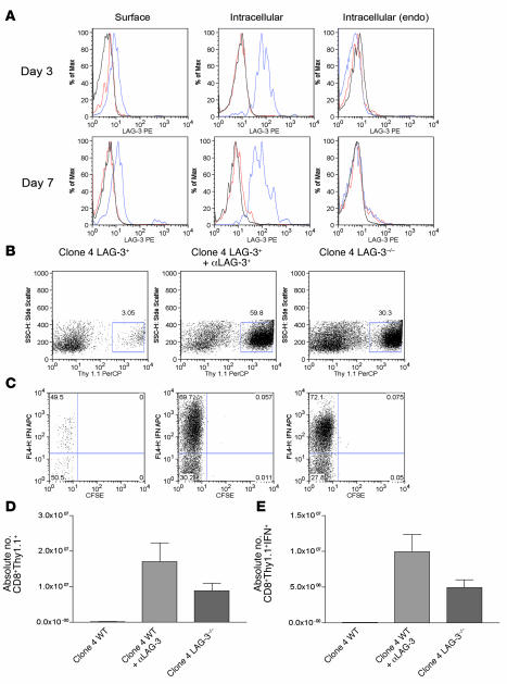
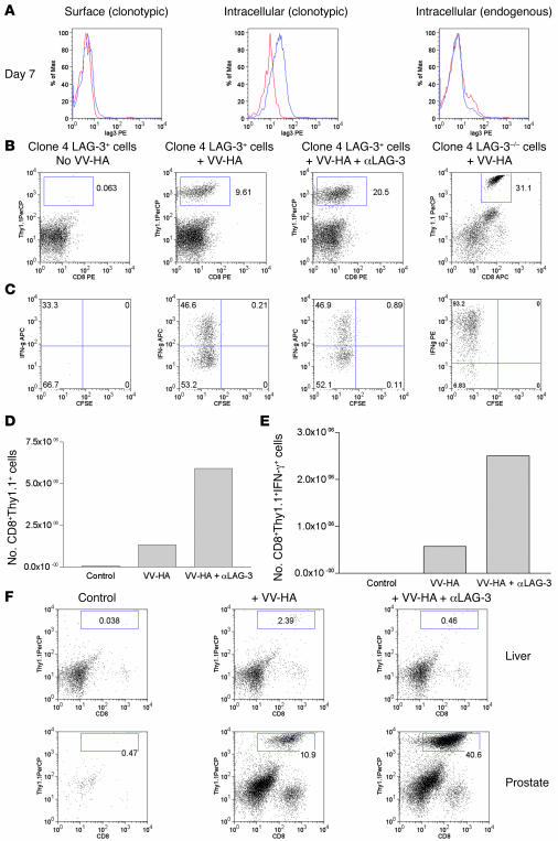
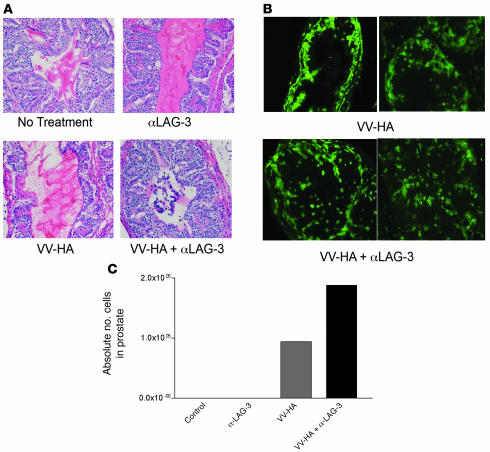
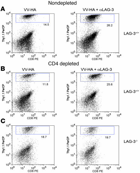
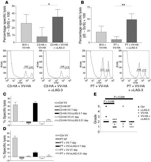
Lymphocyte activation gene-3 (LAG-3) is a cell-surface molecule with diverse biologic effects on T cell function. We recently showed that LAG-3 signaling is important in CD4+ regulatory T cell suppression of autoimmune responses. Here, we demonstrate that LAG-3 maintains tolerance to self and tumor antigens via direct effects on CD8+ T cells using 2 murine systems. Naive CD8+ T cells express low levels of LAG-3, and expression increases upon antigen stimulation. Our data show increased levels of LAG-3 protein on antigen-specific CD8+ T cells within antigen-expressing organs or tumors. In vivo antibody blockade of LAG-3 or genetic ablation of the Lag-3 gene resulted in increased accumulation and effector function of antigen-specific CD8+ T cells within organs and tumors that express their cognate antigen. Most notably, combining LAG-3 blockade with specific antitumor vaccination resulted in a significant increase in activated CD8+ T cells in the tumor and disruption of the tumor parenchyma. A major component of this effect was CD4 independent and required LAG-3 expression by CD8+ T cells. Taken together, these data demonstrate a direct role for LAG-3 on CD8+ T cells and suggest that LAG-3 blockade may be a potential cancer treatment.


Figures

Figure 1
Figure 1. LAG-3 blockade enhances the accumulation of HA-specific CD8+ cells in a model of self tolerance.
We transferred 106 LAG-3+/+ or LAG-3–/–CD8+Thy1.1+ clone 4 CD8+ cells into Thy1.2+C3-HAhigh mice. Mice receiving antibody were given 0.2 mg αLAG-3 i.p. at the time of transfer and 3 days later. At indicated time points after transfer, single-cell suspensions were made from lungs and analyzed for LAG-3 expression and function. (A) CD8+ and Thy1.1+ were gated to determine expression of LAG-3 on CD8+ T cells taken directly from lungs of C3-HA mice. Both surface expression (left) and intracellular staining (middle and right) were performed for LAG-3 expression. Endogenous (endo) CD8 populations within lungs were also assessed. Blue lines, LAG-3; red lines, rat IgG1 isotype; black lines, LAG-3–/– cells. Max, maximum. (B) Lungs from C3-HA mice were analyzed 7 days after transfer for percentage of CD8+Thy1.1+ cells by staining for Thy1.1 (C). Clonotypic cells from B were analyzed for division by dilution of CFSE and IFN-γ production by intracellular staining after stimulation in vitro in the presence of HA peptide plus monensin for 5 hours. SSC-H, side scatter. (D and E) Absolute numbers of clonotypic and IFN-γ+ clonotypic cells/lung after αLAG-3 treatment or transfer of LAG-3–/– clone 4 cells. The mean from 3 mice per group is shown. Each experiment was performed 3 times with data from 1 representative experiment shown.
Figure 2
Figure 2. LAG-3 blockade enhances the accumulation of clone 4 CD8+ effectors within HA-producing prostates of ProHA × TRAMP mice.
(A) In vivo expression of LAG-3 on clonotypic CD8+ T cells. We transferred 2 × 10⁶ Thy1.1+ clone 4 CD8+ cells into VV-HA–injected ProHA × TRAMP mice. Seven days later, prostates were harvested and single-cell suspensions were gated on CD8+ and Thy1.1+ cells and analyzed for expression of LAG-3. Both surface (left) and intracellular staining (middle and right) were performed for detection of LAG-3. Blue line, LAG-3; red line, rat IgG1 isotype; black line, LAG-3–/– cells. (B) αLAG-3 enhances the accumulation of clonotypic cells in prostates. We transferred 106 LAG-3+/+ or LAG-3–/–CD8+Thy1.1+ clone 4 CD8+ cells into ProHA × TRAMP mice. Mice were given VV-HA, VV-HA plus αLAG-3, or nothing at the time of transfer. Mice receiving αLAG-3 were given another dose 3 days later. Seven days after transfer, prostates and livers were collected and homogenized, and single-cell suspensions were analyzed for IFN-γ by intracellular staining after stimulation in vitro in the presence of HA peptide plus monensin for 5 hours. Prostates were analyzed for (B) percentage of CD8+Thy1.1+ and (C) percentage of IFN-γ+ clonotypic cells. Absolute numbers of pooled prostates to determine (D) prostate-derived clonotypic cells and (E) IFN-γ+ clonotypic cells are shown. Error bars are absent because pooling was necessary to count CD8+ cells from prostate tissue. The results are representative of at least 4 experiments. (F) Enhanced accumulation of clone 4 CD8+ cells using αLAG-3 is specific for organs expressing cognate antigen. Livers from ProHA × TRAMP mice do not express the HA antigen (data not shown) and fail to attract and promote division of HA-specific CD8+ T cells. Data from 1 of at least 3 individual experiments are shown. PerCP, peridinin chlorophyll protein.
Figure 3
Figure 3. Visualization of clone 4 cells within prostates and tumor growth inhibition after αLAG-3 treatment.
We transferred 106 Thy1.1+ clone 4 CD8+ cells into 12- to 15-week-old Thy1.2+ ProHA × TRAMP mice. Mice received VV-HA with or without αLAG-3 on the same day as the cell transfers. Mice receiving αLAG-3 were reinjected with αLAG-3 3 days after transfer. Seven days after cell transfers, mice were sacrificed. Prostates from each group (control, αLAG-3, VV-HA, and VV-HA/αLAG-3) were pooled and subsequently split into 2 groups: one for frozen sections (A and B) and the other for flow cytometry analysis (C). Few prostate-infiltrating lymphocytes were seen in nontreated or αLAG-3–treated mice. H&E and Thy1.1 immunofluorescence revealed lymphocytic infiltration after VV-HA and VV-HA/αLAG-3 treatment with disrupted architecture and Thy1.1 cells invading the prostatic lumen only after VV-HA/αLAG-3 treatment. For Thy1.1 staining, no visible cells were present in control or αLAG-3 samples (data not shown). Original magnification, ×200.
Figure 4
Figure 4. Cellular mechanism of αLAG-3 is independent of CD4+ cells but dependent on CD8+ in ProHA × TRAMP prostate tissue.
Intact or GK1.5-treated ProHA × TRAMP mice were injected with VV-HA or VV-HA/αLAG-3 as described and injected with 106 (A and B) clone 4 CD8+ or (C) LAG-3–/– clone 4 CD8+ cells i.v. Mice treated with GK1.5 were bled 1 day before transfer to confirm CD4 depletion. Seven days after cells were transferred, prostates were harvested and single-cell suspensions were prepared as described. Flow cytometry was performed on cells using CD8+ and Thy1.1 antibodies to quantitate percentage of clonotypic cells. Results are representative of 3 experiments.
Figure 5
Figure 5. Self- and tumor-tolerized endogenous CTLs regain effector function in vivo after treatment with VV-HA/αLAG-3.
(A) C3-HA and (B) ProHA × TRAMP mice were given 0.2 mg αLAG-3 i.p. on days 0 and 3 and 106 PFUs of VV-HA i.p. on day 1. Mice received target cells i.v. consisting of a 1:1 mixture of B10.D2 splenocytes loaded with either HA peptide plus 5 mM CFSE or 0.5 mM CFSE only on day 6. Eighteen hours after target transfer, spleens were harvested for flow cytometry. Histograms under each graph represent 1 of 5 individual mice from 1 experiment. Data represent 1 experiment with each being repeated at least 2 times. *P = 0.0378; **P = 0.0171. In vivo CTL assays were performed as above with the addition of groups that were transferred with target cells 21 days after immunization with VV-HA or VV-HA plus αLAG-3 (C and D). (E) Seven-week-old ProHA × TRAMP mice were injected with αLAG-3, VV-HA, or VV-HA/αLAG-3 or left untreated (n = 15 mice/group). Eight weeks later, dorsal prostate lobes were harvested and H&E sections prepared. Slides were graded on a scale from 0–5 as described in Methods in a double-blinded fashion by 2 surgical pathologists. PT, ProHA × TRAMP.

References

    1. Croft M. Co-stimulatory members of the TNFR family: keys to effective T-cell immunity? Nat. Rev. Immunol. . 2003;3:609–620. - PubMed
    1. Dong C., Nurieva R.I., Prasad D.V. Immune regulation by novel costimulatory molecules. Immunol. Res. . 2003;28:39–48. - PubMed
    1. Khoury S.J., Sayegh M.H. The roles of the new negative T cell costimulatory pathways in regulating autoimmunity. Immunity. . 2004;20:529–538. - PubMed
    1. Wang S., Chen L. Co-signaling molecules of the B7-CD28 family in positive and negative regulation of T lymphocyte responses. Microbes Infect. . 2004;6:759–766. - PubMed
    1. Greenwald R.J., Freeman G.J., Sharpe A.H. The B7 family revisited. Annu. Rev. Immunol. . 2005;23:515–548. - PubMed

Publication types

Substances