. 2008 Apr;71(1):320-35.
doi: 10.1002/prot.21679.

Electrostatic rate enhancement and transient complex of protein-protein association

Affiliations

Electrostatic rate enhancement and transient complex of protein-protein association

Ramzi Alsallaq et al. Proteins. 2008 Apr.

Abstract

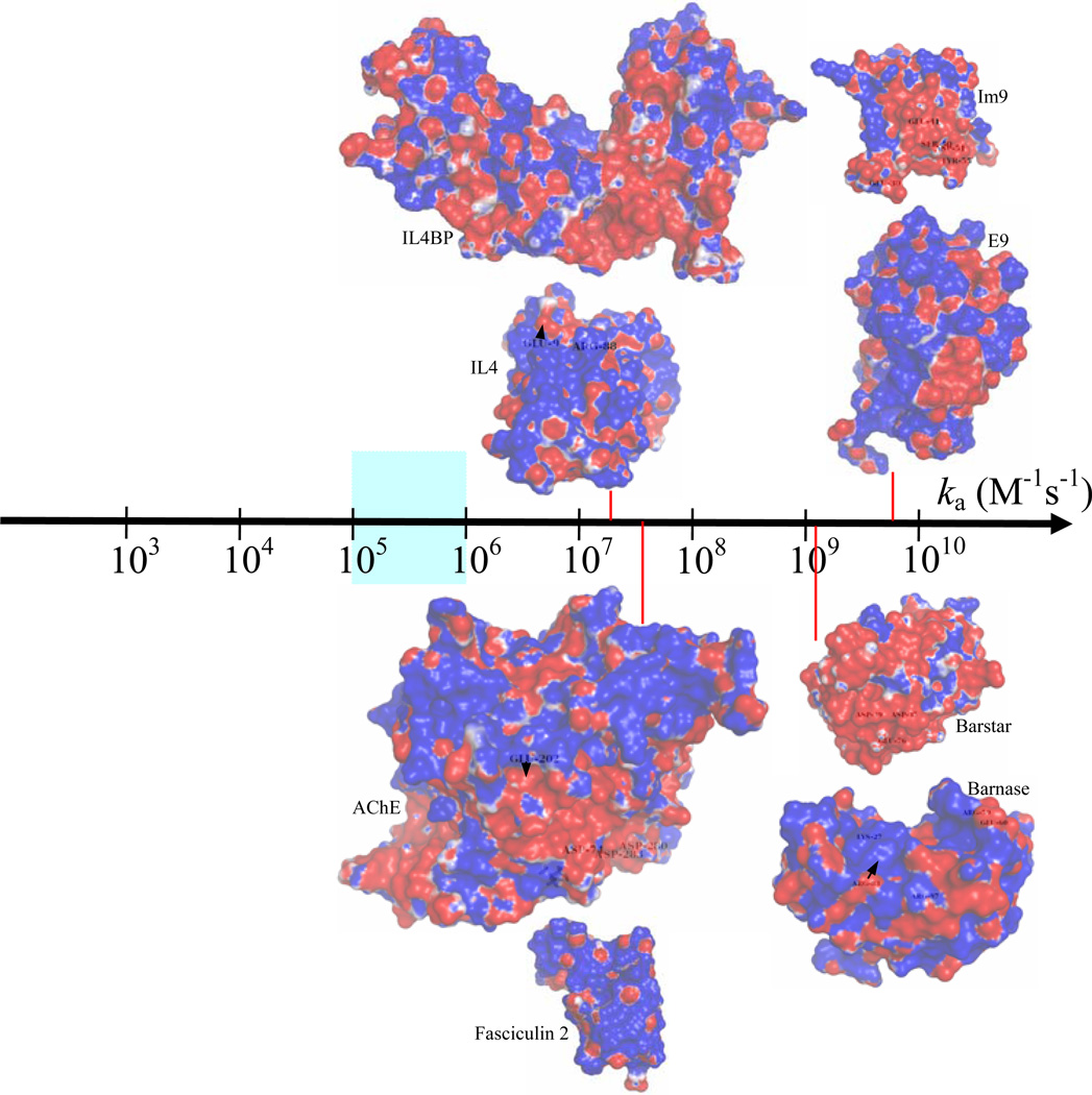
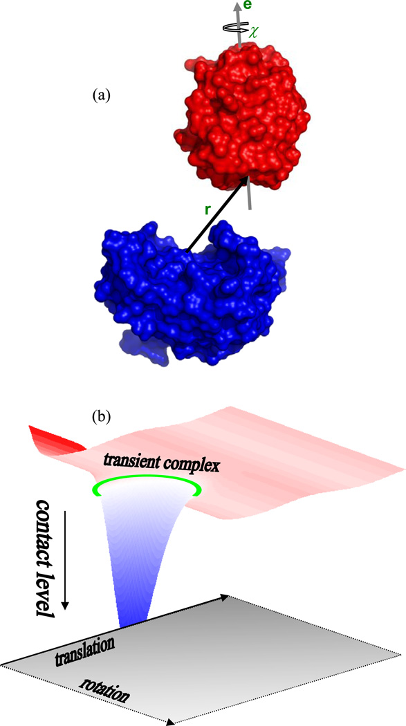
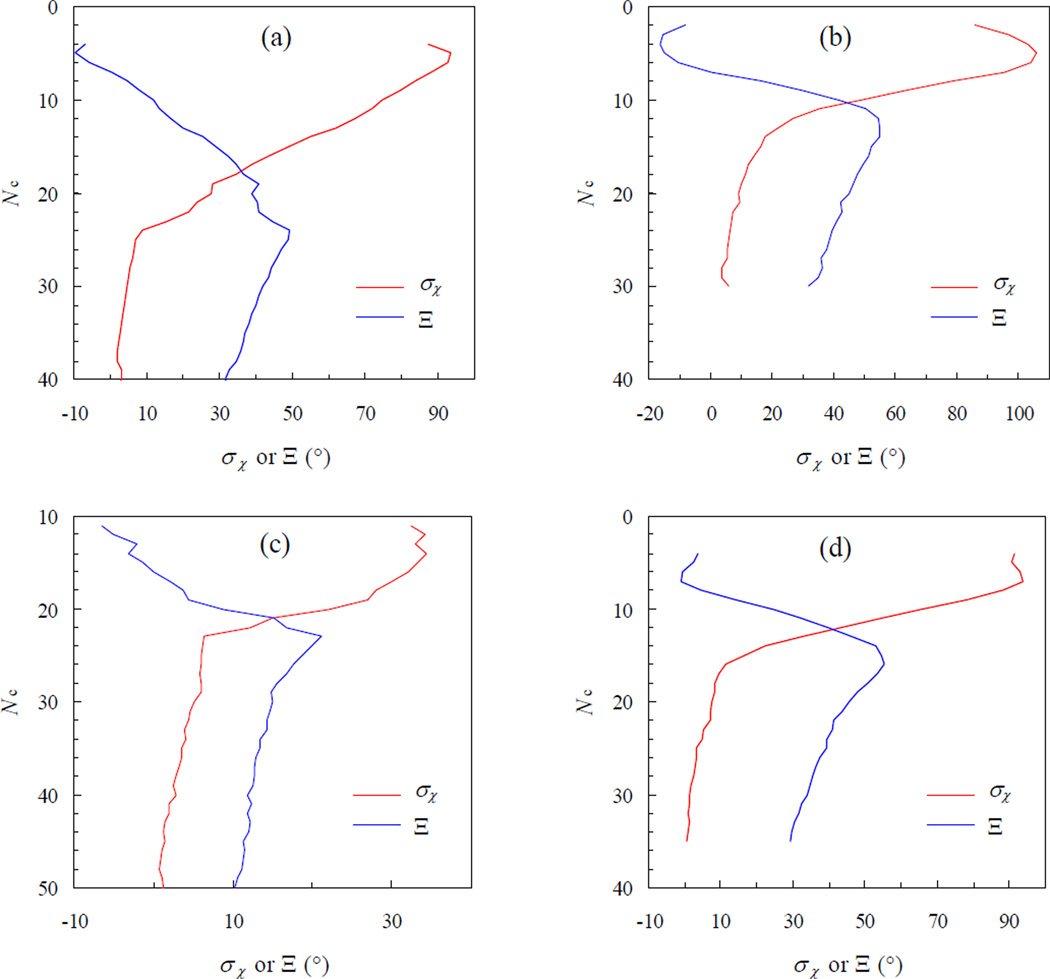
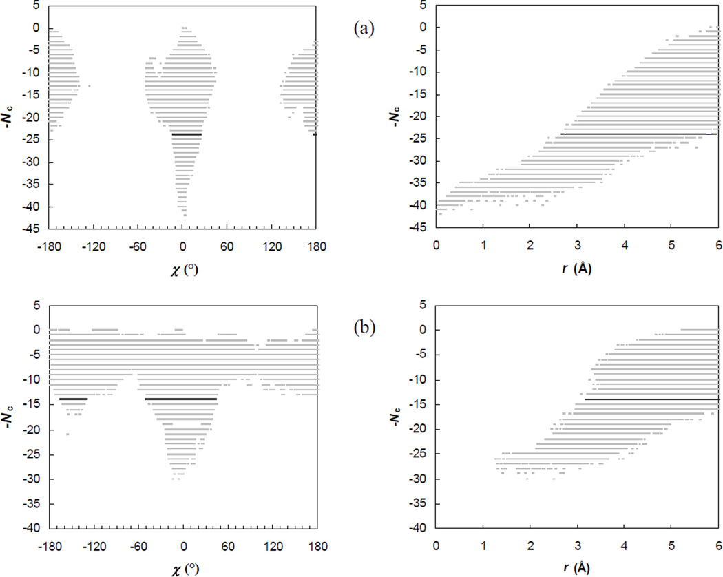
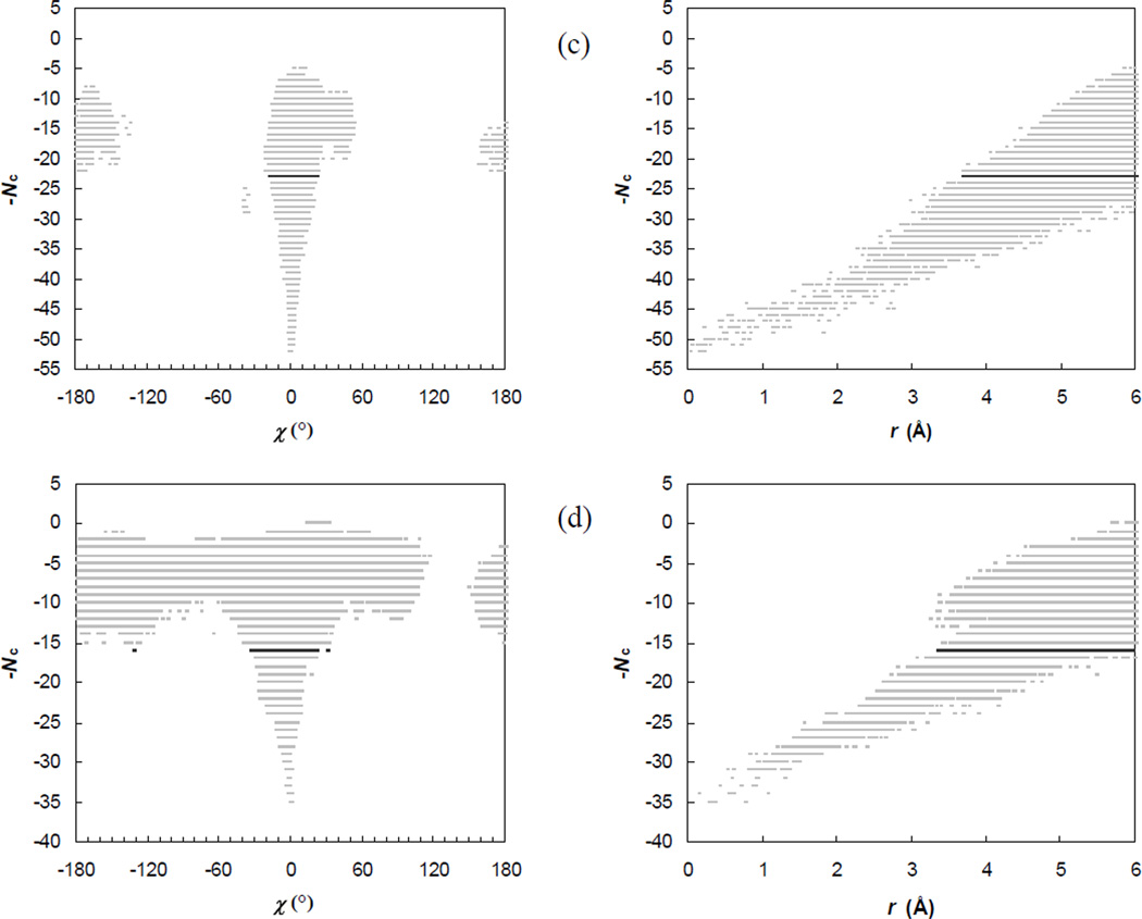
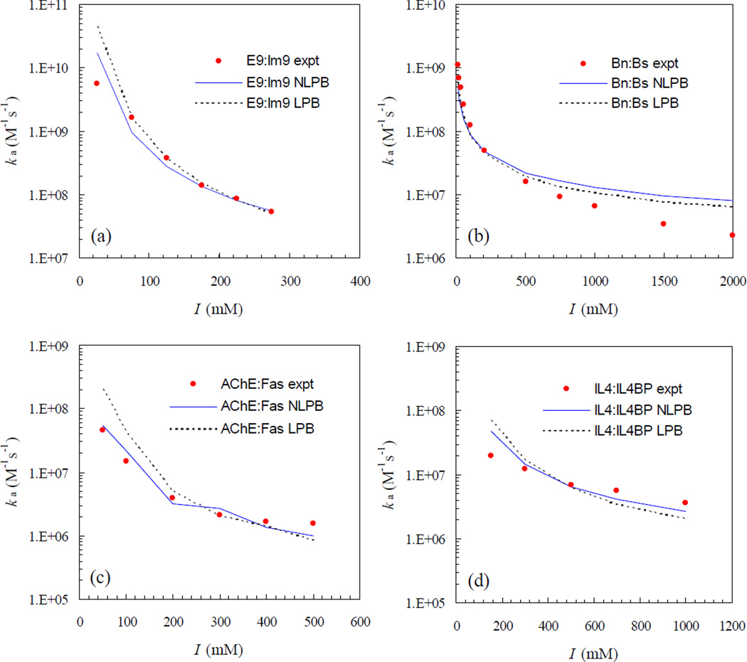
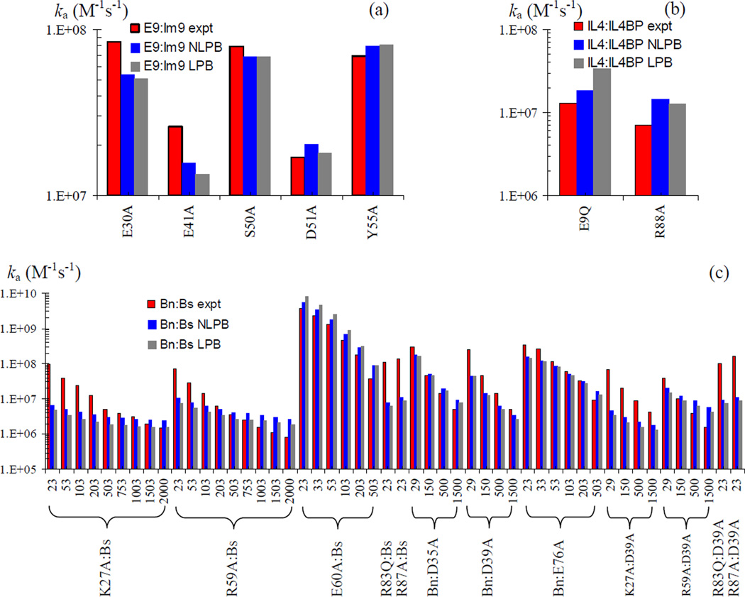
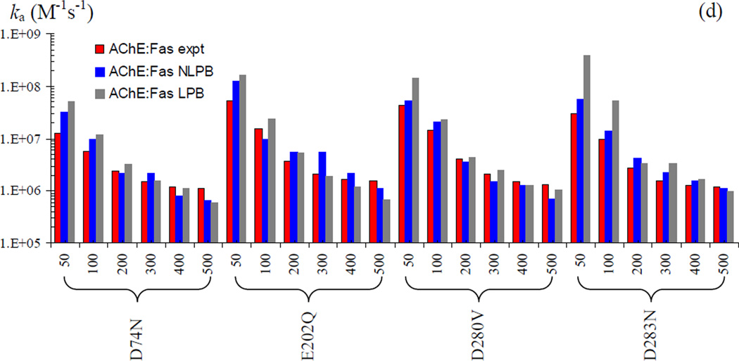
The association of two proteins is bounded by the rate at which they, via diffusion, find each other while in appropriate relative orientations. Orientational constraints restrict this rate to approximately 10(5)-10(6) M(-1) s(-1). Proteins with higher association rates generally have complementary electrostatic surfaces; proteins with lower association rates generally are slowed down by conformational changes upon complex formation. Previous studies (Zhou, Biophys J 1997;73:2441-2445) have shown that electrostatic enhancement of the diffusion-limited association rate can be accurately modeled by $k_{\bf D}$ = $k_{D}0\ {exp} ( - \langle U_{el} \rangle;{\star}/k_{B} T),$ where k(D) and k(D0) are the rates in the presence and absence of electrostatic interactions, respectively, U(el) is the average electrostatic interaction energy in a "transient-complex" ensemble, and k(B)T is the thermal energy. The transient-complex ensemble separates the bound state from the unbound state. Predictions of the transient-complex theory on four protein complexes were found to agree well with the experiment when the electrostatic interaction energy was calculated with the linearized Poisson-Boltzmann (PB) equation (Alsallaq and Zhou, Structure 2007;15:215-224). Here we show that the agreement is further improved when the nonlinear PB equation is used. These predictions are obtained with the dielectric boundary defined as the protein van der Waals surface. When the dielectric boundary is instead specified as the molecular surface, electrostatic interactions in the transient complex become repulsive and are thus predicted to retard association. Together these results demonstrate that the transient-complex theory is predictive of electrostatic rate enhancement and can help parameterize PB calculations.


Figures

Fig. 1
Fig. 1
The wide spectrum of protein-protein association rates. The value 105 M−1s−1 serves as the demarcation point separating the diffusion-limited regime from the conformational change-limited regime. In the diffusion-limited regime, rates in the narrow range of 105 – 106 M−1s−1 are observed for association between proteins, such as antibody and antigen, which do not involve significant electrostatic contributions. Proteins that associate with higher rates typically have complementary electrostatic surfaces, as illustrated by the four protein complexes studied here. The association rates indicated for the E9:Im9, Bn:Bs, AChE:Fas, and IL4:IL4BP complexes are experimental results measured at ionic strengths of 25, 13, 50 and, 150 mM, respectively.,,, Electrostatic potential surfaces are generated by the APBS program and displayed by PyMOL (http://www.pymol.org).
Fig. 2
Fig. 2
(a) Definition of six translational and rotational coordinates for two associating proteins. One protein, shown in blue, is fixed in space; the other, shown in red, can freely translate and rotate. The three translational degrees of freedom are represented by the displacement vector r between the centers of the binding surfaces on the two proteins. Of the three rotational degrees of freedom, two are a unit vector e attached to the moving protein and the remaining one is the rotational angle χ around the unit vector. The unit vector is perpendicular to a plane defined by the binding surface. (b) Illustration of the energy landscape for protein-protein association. The bound state is located in a deep “well” with high levels of contact. The transient-complex ensemble, indicated by a green ring, marks the termination of sharp decrease in contact level and the onset of sharp increase in translational and rotation freedom.
Fig. 3
Fig. 3
Transition of the standard deviation of χ, σχ, from the bound state (with high contact levels) to the unbound state (with low contact levels). The start of the sharp increase in σχ marks the transient complex, with the corresponding contact level Nc* uniquely determined by the maximum of Ξ. Panels (a) – (d) are for the E9:Im9, Bn:Bs, AChE:Fas, and IL4:IL4BP complexes, respectively.
Fig. 4
Fig. 4
Scatter plots of the contact level Nc versus the rotation angle χ or the relative separation r. For clarity, the full range of χ, from −180° to 180° is divided into 500 bins and, at each contact level, at most one sampled χ value is saved for displaying. The sampled configurations are selected in an analogous manner for displaying in the Nc-r scatter plot, with the sampled range of r, from 0 to 6 Å, divided into 500 pins. The Nc-χ and Nc-r pairs of plots labeled (a) – (d) are for the E9:Im9, Bn:Bs, AChE:Fas, and IL4:IL4BP complexes, respectively.
Fig. 4
Fig. 4
Scatter plots of the contact level Nc versus the rotation angle χ or the relative separation r. For clarity, the full range of χ, from −180° to 180° is divided into 500 bins and, at each contact level, at most one sampled χ value is saved for displaying. The sampled configurations are selected in an analogous manner for displaying in the Nc-r scatter plot, with the sampled range of r, from 0 to 6 Å, divided into 500 pins. The Nc-χ and Nc-r pairs of plots labeled (a) – (d) are for the E9:Im9, Bn:Bs, AChE:Fas, and IL4:IL4BP complexes, respectively.
Fig. 5
Fig. 5
Salt dependences of the association rates of the four protein complexes. Experimental results are shown in circles; predictions by the nonlinear (NLPB) and linearized (LPB) Poisson-Boltzmann equations are shown in solid and dotted curves, respectively.
Fig. 6
Fig. 6
Mutation effects on the association rates of the four protein complexes. Experimental and predicted (NLPB and LPB) rates are shown as red, blue, and black bars, respectively. (a) Rates for five Im9 mutations in the E9:Im9 complex at I = 225 mM. (b) Rates for two IL4 mutations in the IL4:IL4BP complex at I = 150 mM. (c) Rates for 12 mutations on the Bn:Bs complex at the various ionic strengths (in mM) indicated. (d) Rates for four AChE mutations in the AChE:Fas complex at the various ionic strengths (in mM) indicated.
Fig. 6
Fig. 6
Mutation effects on the association rates of the four protein complexes. Experimental and predicted (NLPB and LPB) rates are shown as red, blue, and black bars, respectively. (a) Rates for five Im9 mutations in the E9:Im9 complex at I = 225 mM. (b) Rates for two IL4 mutations in the IL4:IL4BP complex at I = 150 mM. (c) Rates for 12 mutations on the Bn:Bs complex at the various ionic strengths (in mM) indicated. (d) Rates for four AChE mutations in the AChE:Fas complex at the various ionic strengths (in mM) indicated.

References

    1. Northrup SH, Erickson HP. Kinetics of protein-protein association explained by Brownian dynamics computer simulation. Proc Natl Acad Sci USA. 1992;89:3338–3342. - PMC - PubMed
    1. Zhou H-X. Enhancement of protein-protein association rate by interaction potential: Accuracy of prediction based on local Boltzmann factor. Biophys J. 1997;73:2441–2445. - PMC - PubMed
    1. Schlosshauer M, Baker D. Realistic protein-protein association rates from a simple diffusional model neglecting long-range interactions, free energy barriers, and landscape ruggedness. Protein Sci. 2004;13(6):1660–1669. - PMC - PubMed
    1. Foote J, Eisen HN. Kinetic and affinity limits on antibodies produced during immune responses. Proc Natl Acad Sci USA. 1995;92(5):1254–1256. - PMC - PubMed
    1. Hoffman TL, LaBranche CC, Zhang W, Canziani G, Robinson J, Chaiken I, Hoxie JA, Doms RW. Stable exposure of the coreceptor-binding site in a CD4-independent HIV-1 envelope protein. Proc Natl Acad Sci USA. 1999;96(11):6359–6364. - PMC - PubMed

Publication types

LinkOut - more resources