. 2007;14(1):13-22.
doi: 10.3727/000000007783991754.

Cloning, expression, and functional analysis of rat liver cytosolic inorganic pyrophosphatase gene and characterization of its functional promoter

Affiliations

Cloning, expression, and functional analysis of rat liver cytosolic inorganic pyrophosphatase gene and characterization of its functional promoter

Harekrushna Panda et al. Gene Expr. 2007.

Abstract

Inorganic pyrophosphate (PPi) is formed in several metabolic processes and its hydrolysis by the ubiquitously expressed enzyme inorganic pyrophosphatase (iPPase) is essential for the reactions to proceed in the direction of biosynthesis. Recently, we have reported differential expression and activity of cytosolic iPPase in rat liver with aging. In this article we report the cloning of the coding region of rat liver cytosolic iPPase gene in a bacterial expression vector, its expression, purification, and functional analysis by in-gel enzyme assay. SDS-PAGE and Western blot analysis of this expressed protein revealed that its molecular weight (MW) is approximately 33 kDa, while in-gel assay showed that it is functionally active just as the liver cytosolic iPPase. We have determined the genomic organization of this gene by genome blast approach. We have also cloned and characterized its proximal approximate 1 kb functional promoter (-1009 to +82) by transient transfection and luciferase assay of different 5'-deleted iPPase promoter-luciferase constructs and also established its transcription start site by primer extension analysis, along with protein-DNA interaction studies for a few putative transcription factor binding sites.


Figures
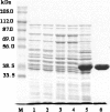
Figure 1
Figure 1
SDS-PAGE analysis of expressed His-tagged rat liver cytosolic inorganic pyrophosphatase. Lane M: molecular weight marker in kDa. Whole cell lysate of: lane 1, E. coli cells; lane 2, 15-h IPTG-induced E. coli cells transformed with self-ligated expression vector pTrcHis TOPO TA ; lane 3, 15-h IPTG-induced E. coli cells transformed with expression vector containing rat liver cytosolic iPPase coding region out-of-frame with the vector; lane 4, 0-h IPTG-induced E. coli cells transformed with expression vector containing rat liver cytosolic iPPase coding region in-frame with the vector; lane 5, 15-h IPTG-induced E. coli cells transformed with expression vector containing rat liver cytosolic iPPase coding region in-frame with the vector; lane 6, Ni-NTA purified recombinant rat liver cytosolic iPPase protein by denaturing method. In each lane 10 μg of protein was taken except for lane 6, where 5 μg purified recombinant protein was used.
Figure 2
Figure 2
Western blot analysis of expressed His-tagged rat liver cytosolic inorganic pyrophosphatase using anti-His-HRP-conjugated antibody. Whole cell lysate of: lane 1, E. coli cells; lane 2, 15-h IPTG-induced E. coli cells transformed with self-ligated expression vector pTrcHis TOPO TA ; lane 3, 15-h IPTG-induced E. coli cells transformed with expression vector containing rat liver cytosolic iPPase coding region out-of-frame with the vector; lane 4, 0-h IPTG-induced E. coli cells transformed with expression vector containing rat liver cytosolic iPPase coding region in-frame with the vector; lane 5, 15-h IPTG-induced E. coli cells transformed with expression vector containing rat liver cytosolic iPPase coding region in-frame with the vector; lane 6, Ni-NTA purified recombinant rat liver cytosolic iPPase protein by denaturing method. In each lane 10 μg of protein was taken except for lane 6, where 5 μg purified recombinant protein was used.
Figure 3
Figure 3
Study of functional activity of expressed rat liver cytosolic iPPase by in-gel assay. Whole cell lysate protein (12 μg) of: lane 1, E. coli cells; lane 2, 15-h IPTG-induced E. coli cells transformed with self-ligated expression vector pTrcHis TOPO TA; lane 3, 15-h IPTG-induced E. coli cells transformed with expression vector containing rat liver cytosolic iPPase coding region out-of-frame with the vector; lane 4, 0-h IPTG-induced E. coli cells transformed with expression vector containing rat liver cytosolic iPPase coding region in-frame with the vector; lane 5, 15-h IPTG-induced E. coli cells transformed with expression vector containing rat liver cytosolic iPPase coding region in-frame with the vector. Lane 6: 5 μg of Ni-NTA column purified renatured rat liver cytosolic iPPase. Lane 7: 40 μg of rat liver WCL. Lanes 1–4 show only endogenous iPPase activity of E. coli. Lane 5 shows activity of expressed rat liver iPPase as well as endogenous E. coli iPPase. Lane 6 shows activity of purified bacterially expressed rat liver iPPase. Lane 7 shows native rat liver iPPase.
Figure 4
Figure 4
Rat liver cytosolic inorganic pyrophosphatase 1009-bp promoter sequence. Nucleotide sequence of iPPase 1009-bp promoter fragment beyond translational start site showing transcription start site (TSS) and putative transcription factor binding sites. This sequence has been deposited in the GenBank with the accession number DQ978330.
Figure 5
Figure 5
Identification of transcription start site of inorganic pyro-phosphatase gene by primer extension analysis. Lanes 1–4, sequencing reactions; lane 5, primer extension product of yeast tRNA; lane 6, primer extension product of liver total RNA. The sequence corresponding to the transcription start site has been marked by a line and is complementary to the sequencing reactions shown in the figure.
Figure 6
Figure 6
Functional analysis of iPPase promoter by luciferase assay. Relative luciferase activity of different iPPase promoter–reporter constructs was obtained by dividing the luciferase activity by the β-galactosidase activity for normalization of transfection efficiency. All the transfections were repeated in triplicate and the results were expressed as the mean ± SD of three independent experiments. The relative luciferase activity for the construct showing highest activity is taken as 100% and all the comparisons are made relative to this.
Figure 7
Figure 7
Protein-DNA interaction studies. Electrophoretic mobility shift assay (EMSA) for Sp1 site (−102 to −82) (A), Sp1 site (−158 to −138) (B), and NF-κB site (−736 to −717) (C) on iPPase promoter. Lane 1, labeled oligonucleotide duplex without nuclear extract; lanes 2–5, labeled oligonucleotide duplex with 10 μg rat liver nuclear extract (RLNE). Lane 2, no competitor DNA; lane 3, 100-fold molar excess of unlabeled homologous self (corresponding to the particular binding site) oligonucleotide duplex; lane 4, 100-fold molar excess of unlabeled consensus oligonucleotide duplex; lane 5, 100-fold molar excess of unlabeled nonspecific oligonucleotide duplex (SRY). C and F denote the positions of protein-DNA complex and free probe, respectively.

Similar articles

Cited by

References

    1. Baeuerle P. A.; Henkel T. Function and activation of NF-kappa B in the immune system. Annu. Rev. Immunol. 12:141–179; 1994. - PubMed
    1. Baeuerle P. A.; Baltimore D. NF-kappa B: Ten years after. Cell 87:13–20; 1996. - PubMed
    1. Barkett M.; Gilmore T. D. Control of apoptosis by Rel/NF-kappaB transcription factors. Oncogene 18:6910–6924; 1999. - PubMed
    1. Davidson A. M.; Halestrap A. P. Inorganic pyrophosphate is located primarily in the mitochondria of hepatocyte and increases in parallel with the decrease in light-scattering induced by gluconeogenic hormones, butyrate and ionophore A23187. Biochem. J. 254:379–384; 1988. - PMC - PubMed
    1. Dusing M. R.; Wiginton D. A. Sp1 is essential for both enhancer-mediated and basal activation of the TATA-less human adenosine deaminase promoter. Nucleic Acids Res. 22:669–677; 1994. - PMC - PubMed

Publication types

MeSH terms