

LigASite--a database of biologically relevant binding sites in proteins with known apo-structures
- PMID: 17933762
- PMCID: PMC2238865
- DOI: 10.1093/nar/gkm839
LigASite--a database of biologically relevant binding sites in proteins with known apo-structures
Abstract
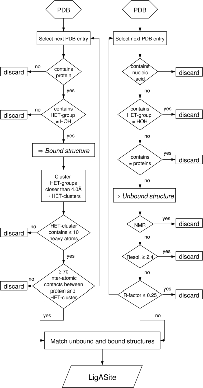
Better characterization of binding sites in proteins and the ability to accurately predict their location and energetic properties are major challenges which, if addressed, would have many valuable practical applications. Unfortunately, reliable benchmark datasets of binding sites in proteins are still sorely lacking. Here, we present LigASite ('LIGand Attachment SITE'), a gold-standard dataset of binding sites in 550 proteins of known structures. LigASite consists exclusively of biologically relevant binding sites in proteins for which at least one apo- and one holo-structure are available. In defining the binding sites for each protein, information from all holo-structures is combined, considering in each case the quaternary structure defined by the PQS server. LigASite is built using simple criteria and is automatically updated as new structures become available in the PDB, thereby guaranteeing optimal data coverage over time. Both a redundant and a culled non-redundant version of the dataset is available at http://www.scmbb.ulb.ac.be/Users/benoit/LigASite. The website interface allows users to search the dataset by PDB identifiers, ligand identifiers, protein names or sequence, and to look for structural matches as defined by the CATH homologous superfamilies. The datasets can be downloaded from the website as Schema-validated XML files or comma-separated flat files.
Figures
References
-
- Rigden DJ. Understanding the cell in terms of structure and function: insights from structural genomics. Curr. Opin. Biotechnol. 2006;17:457–464. - PubMed
-
- Skolnick J, Fetrow JS, Kolinski A. Structural genomics and its importance for gene function analysis. Nat. Biotechnol. 2000;18:283–287. - PubMed
-
- Watson JD, Laskowski RA, Thornton JM. Predicting protein function from sequence and structural data. Curr. Opin. Struct. Biol. 2005;15:275–284. - PubMed
-
- Polacco BJ, Babbitt PC. Automated discovery of 3D motifs for protein function annotation. Bioinformatics. 2006;22:723–730. - PubMed
-
- Jones S, Thornton JM. Searching for functional sites in protein structures. Curr. Opin. Chem. Biol. 2004;8:3–7. - PubMed
