

Analysis of a predicted nuclear localization signal: implications for the intracellular localization and function of the Saccharomyces cerevisiae RNA-binding protein Scp160
- PMID: 17933776
- PMCID: PMC2175298
- DOI: 10.1093/nar/gkm776
Analysis of a predicted nuclear localization signal: implications for the intracellular localization and function of the Saccharomyces cerevisiae RNA-binding protein Scp160
Abstract
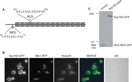
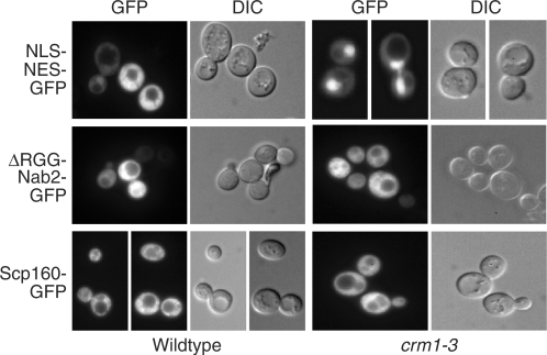
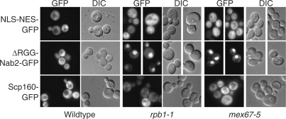
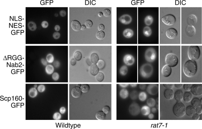
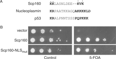
Gene expression is controlled by RNA-binding proteins that modulate the synthesis, processing, transport and stability of various classes of RNA. Some RNA-binding proteins shuttle between the nucleus and cytoplasm and are thought to bind to RNA transcripts in the nucleus and remain bound during translocation to the cytoplasm. One RNA-binding protein that has been hypothesized to function in this manner is the Saccharomyces cerevisiae Scp160 protein. Although the steady-state localization of Scp160 is cytoplasmic, previous studies have identified putative nuclear localization (NLS) and nuclear export (NES) signals. The goal of this study was to test the hypothesis that Scp160 is a nucleocytoplasmic shuttling protein. We exploited a variety of yeast export mutants to capture any potential nuclear accumulation of Scp160 and found no evidence that Scp160 enters the nucleus. These localization studies were complemented by a mutational analysis of the predicted NLS. Results indicate that key basic residues within the predicted NLS of Scp160 can be altered without severely affecting Scp160 function. This finding has important implications for understanding the function of Scp160, which is likely limited to the cytoplasm. Additionally, our results provide strong evidence that the presence of a predicted nuclear localization signal within the sequence of a protein should not lead to the assumption that the protein enters the nucleus in the absence of additional experimental evidence.
Figures
Similar articles
-
Nucleocytoplasmic shuttling of Ssd1 defines the destiny of its bound mRNAs.Mol Microbiol. 2011 Aug;81(3):831-49. doi: 10.1111/j.1365-2958.2011.07731.x. Epub 2011 Jul 18. Mol Microbiol. 2011. PMID: 21762218 Free PMC article.
-
Both KH and non-KH domain sequences are required for polyribosome association of Scp160p in yeast.Nucleic Acids Res. 2004 Sep 8;32(16):4768-75. doi: 10.1093/nar/gkh812. Print 2004. Nucleic Acids Res. 2004. PMID: 15356294 Free PMC article.
-
Nuclear shuttling of She2p couples ASH1 mRNA localization to its translational repression by recruiting Loc1p and Puf6p.Mol Biol Cell. 2009 Apr;20(8):2265-75. doi: 10.1091/mbc.e08-11-1151. Epub 2009 Feb 25. Mol Biol Cell. 2009. PMID: 19244342 Free PMC article.
-
Gatekeepers of the nucleus.Science. 2000 May 26;288(5470):1374-7. doi: 10.1126/science.288.5470.1374. Science. 2000. PMID: 10827939 Review.
-
Nucleocytoplasmic transport enters the atomic age.Curr Opin Cell Biol. 2001 Jun;13(3):310-9. doi: 10.1016/s0955-0674(00)00213-1. Curr Opin Cell Biol. 2001. PMID: 11343901 Review.
Cited by
-
Recruitment, Duplex Unwinding and Protein-Mediated Inhibition of the Dead-Box RNA Helicase Dbp2 at Actively Transcribed Chromatin.J Mol Biol. 2016 Mar 27;428(6):1091-1106. doi: 10.1016/j.jmb.2016.02.005. Epub 2016 Feb 11. J Mol Biol. 2016. PMID: 26876600 Free PMC article.
-
Proteasome-mediated processing of Def1, a critical step in the cellular response to transcription stress.Cell. 2013 Aug 29;154(5):983-995. doi: 10.1016/j.cell.2013.07.028. Cell. 2013. PMID: 23993092 Free PMC article.
References
-
- Tran EJ, Wente SR. Dynamic nuclear pore complexes: life on the edge. Cell. 2006;125:1041–1053. - PubMed
-
- Suntharalingam M, Wente SR. Peering through the pore. Nuclear pore complex structure, assembly, and function. Dev. Cell. 2003;4:775–789. - PubMed
-
- Lim RY, Fahrenkrog B. The nuclear pore complex up close. Curr. Opin. Cell Biol. 2006;18:342–347. - PubMed
-
- Kalderon D, Richardson WD, Markham AF, Smith AE. Sequence requirements for nuclear location of simian virus 40 large-T antigen. Nature. 1984;311:33–38. - PubMed
Publication types
MeSH terms
Substances
Grants and funding
LinkOut - more resources
Full Text Sources
Molecular Biology Databases
