Visualization of detergent solubilization of membranes: implications for the isolation of rafts
- PMID: 17933878
- PMCID: PMC2212709
- DOI: 10.1529/biophysj.107.114108
Visualization of detergent solubilization of membranes: implications for the isolation of rafts
Abstract
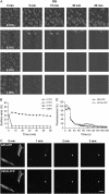
Although different detergents can give rise to detergent-resistant membranes of different composition, it is unclear whether this represents domain heterogeneity in the original membrane. We compared the mechanism of action of five detergents on supported lipid bilayers composed of equimolar sphingomyelin, cholesterol, and dioleoylphosphatidylcholine imaged by atomic force microscopy, and on raft and nonraft marker proteins in live cells imaged by confocal microscopy. There was a marked correlation between the detergent solubilization of the cell membrane and that of the supported lipid bilayers. In both systems Triton X-100 and CHAPS (3-[(3-cholamidopropyl)dimethylammonio]-1-propanesulfonate) distinguished between the nonraft liquid-disordered (l(d)) and raft liquid ordered (l(o)) lipid phases by selectively solubilizing the l(d) phase. A higher concentration of Lubrol was required, and not all the l(d) phase was solubilized. The solubilization by Brij 96 occurred by a two-stage mechanism that initially resulted in the solubilization of some l(d) phase and then progressed to the solubilization of both l(d) and l(o) phases simultaneously. Octyl glucoside simultaneously solubilized both l(o) and l(d) phases. These data show that the mechanism of membrane solubilization is unique to an individual detergent. Our observations have significant implications for using different detergents to isolate membrane rafts from biological systems.
Figures


Similar articles
-
Sphingomyelin chain length influences the distribution of GPI-anchored proteins in rafts in supported lipid bilayers.Mol Membr Biol. 2007 May-Jun;24(3):233-42. doi: 10.1080/09687860601127770. Mol Membr Biol. 2007. PMID: 17520480
-
Differential solubilization of membrane lipids by detergents: coenrichment of the sheep brain serotonin 5-HT1A receptor with phospholipids containing predominantly saturated fatty acids.Arch Biochem Biophys. 1993 Aug 15;305(1):68-77. doi: 10.1006/abbi.1993.1394. Arch Biochem Biophys. 1993. PMID: 8342957
-
Transbilayer peptide sorting between raft and nonraft bilayers: comparisons of detergent extraction and confocal microscopy.Biophys J. 2005 Aug;89(2):1102-8. doi: 10.1529/biophysj.105.062380. Epub 2005 May 20. Biophys J. 2005. PMID: 15908585 Free PMC article.
-
Model systems, lipid rafts, and cell membranes.Annu Rev Biophys Biomol Struct. 2004;33:269-95. doi: 10.1146/annurev.biophys.32.110601.141803. Annu Rev Biophys Biomol Struct. 2004. PMID: 15139814 Review.
-
Detergent solubilization of lipid bilayers: a balance of driving forces.Trends Biochem Sci. 2013 Feb;38(2):85-93. doi: 10.1016/j.tibs.2012.11.005. Epub 2013 Jan 4. Trends Biochem Sci. 2013. PMID: 23290685 Review.
Cited by
-
Antibody library screens using detergent-solubilized mammalian cell lysates as antigen sources.Protein Eng Des Sel. 2010 Jul;23(7):567-77. doi: 10.1093/protein/gzq029. Epub 2010 May 23. Protein Eng Des Sel. 2010. PMID: 20498037 Free PMC article.
-
Direct visualization of the action of Triton X-100 on giant vesicles of erythrocyte membrane lipids.Biophys J. 2014 Jun 3;106(11):2417-25. doi: 10.1016/j.bpj.2014.04.039. Biophys J. 2014. PMID: 24896120 Free PMC article.
-
The Role of Lipid Environment in Ganglioside GM1-Induced Amyloid β Aggregation.Membranes (Basel). 2020 Sep 9;10(9):226. doi: 10.3390/membranes10090226. Membranes (Basel). 2020. PMID: 32916822 Free PMC article. Review.
-
Ceramide signaling in cancer and stem cells.Future Lipidol. 2008 Jun;3(3):273-300. doi: 10.2217/17460875.3.3.273. Future Lipidol. 2008. PMID: 19050750 Free PMC article.
-
Transmembrane adaptor protein PAG/CBP is involved in both positive and negative regulation of mast cell signaling.Mol Cell Biol. 2014 Dec 1;34(23):4285-300. doi: 10.1128/MCB.00983-14. Epub 2014 Sep 22. Mol Cell Biol. 2014. PMID: 25246632 Free PMC article.
References
-
- Pike, L. J. 2006. Rafts defined: a report on the Keystone symposium on lipid rafts and cell function. J. Lipid Res. 47:1597–1598. - PubMed
-
- Jacobson, K., O. G. Mouritsen, and R. G. Anderson. 2007. Lipid rafts: at a crossroad between cell biology and physics. Nat. Cell Biol. 9:7–14. - PubMed
-
- Hooper, N. M. 1999. Detergent-insoluble glycosphingolipid/cholesterol-rich membrane domains, lipid rafts and caveolae. Mol. Membr. Biol. 16:145–156. - PubMed
-
- Simons, K., and D. Toomre. 2000. Lipid rafts and signal transduction. Nat. Rev. Mol. Cell Biol. 1:31–39. - PubMed
-
- Brown, D. A., and E. London. 1998. Structure and origin of ordered lipid domains in biological membranes. J. Membr. Biol. 164:103–114. - PubMed
Publication types
MeSH terms
Substances
Grants and funding
LinkOut - more resources
Full Text Sources
Other Literature Sources
Research Materials

