. 2007 Oct;19(10):3127-45.
doi: 10.1105/tpc.106.048868. Epub 2007 Oct 12.

Subcellular localization and functional domain studies of DEFECTIVE KERNEL1 in maize and Arabidopsis suggest a model for aleurone cell fate specification involving CRINKLY4 and SUPERNUMERARY ALEURONE LAYER1

Affiliations

Subcellular localization and functional domain studies of DEFECTIVE KERNEL1 in maize and Arabidopsis suggest a model for aleurone cell fate specification involving CRINKLY4 and SUPERNUMERARY ALEURONE LAYER1

Qing Tian et al. Plant Cell. 2007 Oct.

Abstract

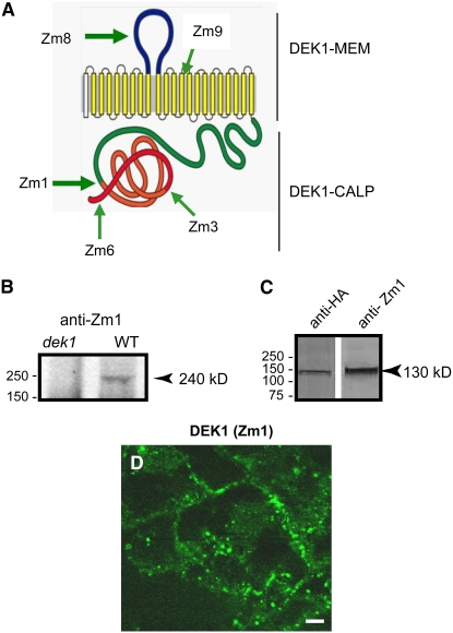
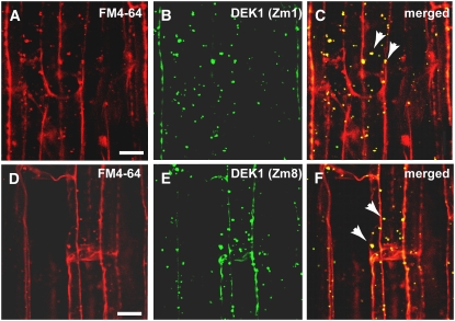
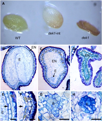
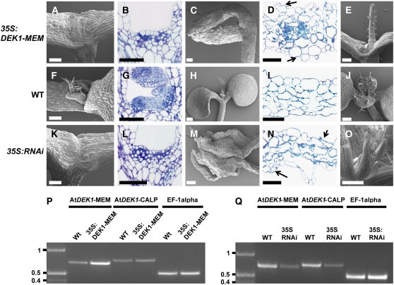
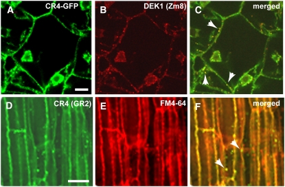
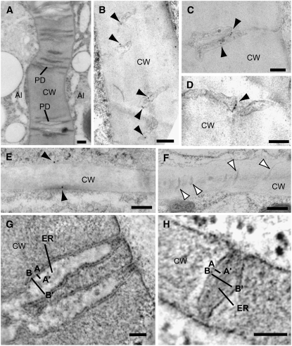
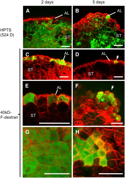
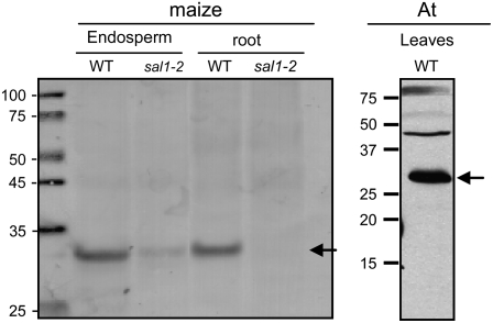
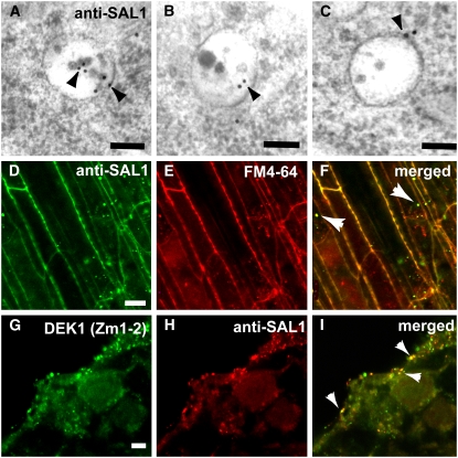
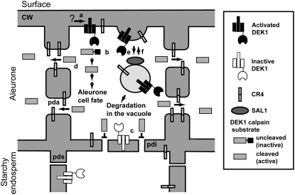
DEFECTIVE KERNEL1 (DEK1), which consists of a membrane-spanning region (DEK1-MEM) and a calpain-like Cys proteinase region (DEK1-CALP), is essential for aleurone cell formation at the surface of maize (Zea mays) endosperm. Immunolocalization and FM4-64 dye incubation experiments showed that DEK1 and CRINKLY4 (CR4), a receptor kinase implicated in aleurone cell fate specification, colocalized to plasma membrane and endosomes. SUPERNUMERARY ALEURONE LAYER1 (SAL1), a negative regulator of aleurone cell fate encoding a class E vacuolar sorting protein, colocalized with DEK1 and CR4 in endosomes. Immunogold localization, dual-axis electron tomography, and diffusion of fluorescent dye tracers showed that young aleurone cells established symplastic subdomains through plasmodesmata of larger dimensions than those connecting starchy endosperm cells and that CR4 preferentially associated with plasmodesmata between aleurone cells. Genetic complementation experiments showed that DEK1-CALP failed to restore wild-type phenotypes in maize and Arabidopsis thaliana dek1 mutants, and DEK1-MEM also failed to restore wild-type phenotypes in Arabidopsis dek1-1 mutants. Instead, ectopic expression of DEK1-MEM under the control of the cauliflower mosaic virus 35S promoter gave a dominant negative phenotype. These data suggest a model for aleurone cell fate specification in which DEK1 perceives and/or transmits a positional signal, CR4 promotes the lateral movement of aleurone signaling molecules between aleurone cells, and SAL1 maintains the proper plasma membrane concentration of DEK1 and CR4 proteins via endosome-mediated recycling/degradation.


Figures

Figure 1.
Figure 1.
Zm DEK1 Antibodies and Subcellular Localization. (A) Zm DEK1 is predicted to consist of 21 membrane-spanning segments embedded in the plasma membrane and an external loop region (DEK1-MEM). The first (unfilled) membrane-spanning segment represents a predicted signal peptide. The cytoplasmic portion harbors a calpain-like Cys proteinase (DEK1-CALP). Polyclonal antibodies were produced against peptides in the regions marked by Zm1, Zm3, Zm6, Zm8, and Zm9. (B) Immunoblot of protein extracts from wild-type and dek1/dek1 kernels at 12 DAP probed with DEK1 Zm1 antibody. The arrowhead indicates the band with the expected size (240 kD) of the DEK1 protein. (C) Immunoblots of protein extracts from Zm Dek1-CALP:HA:FLAG:AcGFP transgenic endosperm probed with anti-HA and DEK1 Zm1 antibodies. The arrowhead indicates a band with the expected size of the fusion protein. (D) Immunostaining of DEK1 using DEK1 Zm1 as the primary antibody and a FITC-conjugated secondary antibody. Bar = 10 μm.
Figure 2.
Figure 2.
Colocalization of DEK1 and FM4-64 in Endocytotic Compartments. (A) to (C) Confocal microscopy images of maize root cells stained with FM4-64 (A) and with the DEK1 Zm1 antibody and a secondary antibody conjugated with FITC (B). (C) shows a merged image of (A) and (B). Yellow indicates overlapping fluorescent signal (arrowheads). (D) to (F) Confocal microscopy images of root cells stained with the endocytic marker FM4-64 (D) and DEK1 Zm8 antibody with a FITC-conjugated secondary antibody (E). (F) shows a merged image of (D) and (E). Yellow indicates overlapping fluorescent signal (arrowheads). Note the colocalization between DEK1 antibodies and FM4-64 in punctate cytoplasmic structures, likely endosomes (arrowheads) Bars = 5 μm.
Figure 3.
Figure 3.
The DEK1 Calpain Cys Proteinase Domain Alone Is Unable to Fully Complement the dek1 Mutant Phenotype in Arabidopsis. (A) Arabidopsis seeds from a DEK1/dek1-1 plant transformed with the At DEK1-CALP expression cassette under the control of the At DEK1 promoter, showing segregation of a wild-type seed (WT), an intermediate phenotype seed (dek1-int), and a collapsed dek1-1 seed (dek1). (B) to (D) Longitudinal sections of Arabidopsis seed types shown in (A): wild-type seed (B); intermediate seed type (C); and typical collapsed dek1-1 seed (D). (E) Detail from a wild-type seed with a peripheral aleurone layer (arrow). (F) Detail from an intermediate seed type lacking aleurone differentiation in the peripheral position of the endosperm (arrow). (G) An embryo from intermediate type seeds. (H) A typical dek1-1 embryo. E, embryo; EN, endosperm; SC, seed coat. Bars = 50 μm.
Figure 4.
Figure 4.
Phenotypic Analysis of Arabidopsis Lines Expressing At DEK1-MEM and At DEK1-RNAi. (A) to (E) Phenotypes of At DEK1-MEM seedlings. (F) to (J) Comparable developmental stages and organs in wild-type seedlings. (K) to (O) Comparable developmental stages and organs in At DEK1-RNAi seedlings. The shoot apex is severely affected in At DEK1-MEM plants ([A] and [B]) and severely affected in RNAi lines ([K] and [L]). Cotyledons from At DEK1-MEM (C) and At DEK1-RNAi (M) seedlings are also distorted. Note in the cross sections of cotyledons from At DEK1-MEM (D) and At DEK1-RNAi (N) plants that the cells exposed to both adaxial and abaxial surfaces contain chloroplasts, suggesting that they have not completely differentiated into epidermal cells. Shoot apices from less severely affected seedlings in both At DEK1-MEM and At DEK1-RNAi lines can display one terminal radialized rosette leaf (E) or several radial rosette leaves (O). Bars = 100 μm. (P) and (Q) RT-PCR analyses showing At DEK1 transcripts in At DEK1-MEM and wild-type seedlings (P) and in At DEK1-RNAi and wild-type seedlings (Q). The three primer sets used, specific for the At DEK1-MEM region, the At DEK1-CALP region, and EF-1α as a control to monitor template presence, are indicated at top. DNA marker sizes are indicated at left in kb.
Figure 5.
Figure 5.
CR4 Antibodies Recognize Zm CR4 in Wild-Type and Transgenic Maize Seeds. (A) Domain structure and location of peptides used to generate Zm CR4 antibodies. (B) Immunoblots of protein extracts from wild-type and cr4 mutant maize roots probed with CR4 GR2 and GR8 antibodies. Arrowheads indicate the band with the expected size of the CR4 protein (∼95 kD). (C) Immunoblots using protein extracts from CR4:HA:FLAG:AcGFP transgenic endosperms (+) or nontransgenic endosperm controls (−) probed with GFP and CR4 antibodies. The arrowhead indicates the fusion protein at 127 kD.
Figure 6.
Figure 6.
Colocalization of CR4 and DEK1 in Endosomes. (A) to (C) Colocalization of CR4 (A) and DEK1 (B) in in vitro–grown CR4:HA:FLAG:AcGFP transgenic maize endosperm. (C) shows a merged image of (A) and (B). The GFP signal and the DEK1 signal overlapped on plasma membrane and punctate structures in the cytoplasm (arrowheads). Bar = 20 μm. (D) to (F) Wild-type maize root sections immunostained with GR2 antibody and a secondary antibody conjugated with FITC (D). (E) shows staining with FM-4-64. (F) shows the two signals colocalized on plasma membrane and in punctate structures in the cytoplasm, likely endosomes (arrowheads). Bar = 10 μm.
Figure 7.
Figure 7.
Electron Microscopy Analysis of CR4 Localization in Maize. (A) General overview of branched plasmodesmata (PD) connecting aleurone cells in a high-pressure frozen/freeze-substituted in vitro–grown maize endosperm. Al, aleurone; CW, cell wall. (B) to (D) Immunogold labeling of CR4 (GR2 antibodies) on plasmodesmata (arrowheads) between aleurone cells. (E) and (F) Immunogold labeling of CR4 on the plasma membrane (black arrowheads in [E]) of starchy endosperm cells. The plasmodesmata connecting starchy endosperm cells (white arrowheads in [F]) are much narrower than those located between aleurone cells and showed very low or no CR4 labeling. (G) and (H) Tomographic slices of plasmodesmata in cell walls between aleurone cells (G) and between starchy endosperm cells (H). Line A to A′ indicates the distance between the plasma membrane and the endoplasmic reticulum membrane (ER), and line B to B′ indicates the widest total diameter of the plasmodesmata. Bars = 100 nm in (A) to (F) and 50 nm (G) and (H).
Figure 8.
Figure 8.
Cell-to-Cell Movement of Fluorescent Tracers in in Vitro–Grown Maize Endosperms. (A) and (B) Endosperms grown in vitro for 2 d (A) or 5 d (B) allow the movement of HPTS through both differentiating aleurone (AL) and starchy endosperm (ST) cells. (C) to (H) In vitro–grown endosperm incubated in 40 kD of F-dextran. The tracer is able to move only through the aleurone cells of endosperms cultured for 2 d ([C], [E], and [G]) or 5 d ([D], [F], and [H]), but the movement is highly reduced in the latter. Most of the aleurone cells that contain the 40-kD F-dextran in the 5-d-in-culture endosperms are located at growing bulges at the endosperm surface ([F], arrow). (G) and (H) show paradermal views of the aleurone layer in endosperms grown in vitro for 2 d (G) or 5 d (H). Note the reduction in the aleurone cells containing the 40-kD F-dextran tracer in the older endosperms. Bars = 100 μm.
Figure 9.
Figure 9.
Characterization of SAL1 Antibodies. Immunoblots of protein extracts from maize wild type and sal1-2 mutant endosperms and roots as well as Arabidopsis leaf proteins (At) probed with anti-SAL1 antibodies. Arrows show bands at the predicted size for SAL1. The presence of a weak band in the mutant shows that the maize sal1-2 mutant is not a null allele.
Figure 10.
Figure 10.
Localization of SAL1 in Arabidopsis and Maize Cells. (A) to (C) Immunogold labeling of multivesicular endosomes in high-pressure frozen/freeze-substituted Arabidopsis embryo cells with anti-SAL1 antibodies. The antibodies labeled 60% of the multivesicular bodies analyzed (n = 40). (D) to (F) Colocalization of SAL1 and the endocytotic marker FM4-64 in punctate structures, likely endosomes (arrowheads in [F]), in wild-type maize roots. (D) shows immunolabeling with anti-SAL1 antibodies and a FITC-conjugated secondary antibody, and (E) shows staining with fixable FM4-64. (F) shows a merged image of (D) and (E). (G) to (I) In vitro endosperm section immunostained with DEK1 rat antibody Zm1-2 (G) and SAL1 (H). Overlapping staining (I) is indicated by arrowheads in the merged image of (G) and (H). Bars = 10 μm.
Figure 11.
Figure 11.
A Model for the Role of DEK1, CR4, and SAL1 in Aleurone Cell Specification. DEK1 at the surface of the endosperm is activated by an unknown mechanism (a), its calpain domain in the cytosol cleaving a postulated substrate (b) that leads to the specification of aleurone cell fate. DEK1 in all other positions is inactive (c). In cells with active DEK1 signaling, CR4 concentrates on plasmodesmata between aleurone cells (pda) and increases the plasmodesma exclusion limit, allowing the activated DEK1 substrate to move laterally between aleurone cells, thereby reinforcing the signal for aleurone cell fate specification (d). Plasmodesmata in cell walls between starchy endosperm cells are narrow (pds), whereas plasmodesmata in cell walls between aleurone cells and starchy endosperm cells are intermediary in width (pdi). DEK1 and CR4 are internalized by endocytosis (e) and traffic through endosomes. Whereas some DEK1 and CR4 molecules may be recycled back to the plasma membrane (f), others are sorted for degradation in the vacuole in a process that requires SAL1. Some endosomes are recycled back to the plasma membrane (f).

Similar articles

Cited by

References

    1. Aggarwal, B.B. (2003). Signalling pathways of the TNF superfamily: A double-edged sword. Nat. Rev. Immunol. 3 745–756. - PubMed
    1. Babst, M. (2005). A protein's final ESCRT. Traffic 6 2–9. - PubMed
    1. Becraft, P.W., and Asuncion-Crabb, Y. (2000). Positional cues specify and maintain aleurone cell fate in maize endosperm development. Development 127 4039–4048. - PubMed
    1. Becraft, P.W., Kang, S.-H., and Suh, S.-G. (2001). The maize CRINKLY4 receptor kinase controls a cell-autonomous differentiation response. Plant Physiol. 127 486–496. - PMC - PubMed
    1. Becraft, P.W., Li, K., Dey, N., and Asuncion-Crabb, Y. (2002). The maize Dek1 gene functions in embryonic pattern formation and cell fate specification. Development 129 5217–5225. - PubMed

Publication types

MeSH terms