. 2007 Dec;6(12):2240-50.
doi: 10.1128/EC.00104-07. Epub 2007 Oct 12.

A Rho3 homolog is essential for appressorium development and pathogenicity of Magnaporthe grisea

Affiliations

A Rho3 homolog is essential for appressorium development and pathogenicity of Magnaporthe grisea

Wu Zheng et al. Eukaryot Cell. 2007 Dec.

Abstract

The small GTPase Rho3 is conserved in fungi and plays a key role in the control of cell polarity and exocytosis in yeast. In this report, we show that a Rho3 homolog, MgRho3, is dispensable for polarized hyphal growth in the rice blast fungus Magnaporthe grisea. However, MgRho3 is required for plant infection. Appressoria formed by the Mgrho3 deletion mutants are morphologically abnormal and defective in plant penetration. Conidia of the Mgrho3 deletion mutants are narrower than those of the wild-type strain and delayed in germination. Transformants expressing a dominant negative Mgrho3 allele exhibit similar phenotypes as the Mgrho3 deletion mutant, while transformants expressing a constitutively active allele of MgRho3 can produce normal conidia but remain defective in appressorium formation and plant infection. In contrast, overexpression of wild-type MgRho3 increases the infectivity of M. grisea. Our results reveal a new role for the conserved Rho3 as a critical regulator of developmental processes and pathogenicity of M. grisea.


Figures
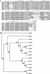
FIG. 1.
FIG. 1.
Alignment and phylogenetic analysis of MgRho3 with other fungal Rho3 homologs. (A) An alignment of Rho3 homologs in fungi based on their amino acid sequences. Five GTP/GDP binding or hydrolysis domains are highlighted and labeled as G1 through G5. G1, GXXXXGKS/T; G2, core effector domain T; G3, DXXGQ/H/T; G4, T/NKXD; G5, C/SAK/L/T); CAAX box, a posttranslational prenylation motif. (B) Phylogenetic relationship of Rho3 homologs in fungi, calculated using the neighbor-joining method of parsimony distance in PHYLIP 3.65 (bootstrap values [percentages] are indicated at the nodes). The fungal source and accession number of each sequence are represented as follows: AfRho3, Aspergillus fumigatus, AAG12157; AnRho3, Aspergillus nidulans, XP_660291; CaRho3, Candida albicans, EAK95917; EgRho3, Eremothecium gossypii, AAG41252; EnRho3, Emericella nidulans, AAK55443; FgRho3, Fusarium graminearum, XP_380346; HjRho3, Hypocrea jecorina, CAC20376; MgRho3, Magnaporthe grisea, MG07176.4); NcRho3, Neurospora crassa, CAE76595; ScRho1, Saccharomyces cerevisiae, BAA00897; SccRho3, Schizophyllum commune, Q9P8J9; SpRho1, Schizosaccharomyces pombe, O13928; UmRho3, Ustilago maydis, XP_401685. The tree was rooted with AtROP1 (Arabidopsis thaliana, NP190698) as an outgroup member.
FIG. 2.
FIG. 2.
The MgRho3 knockout construct and molecular confirmation. (A) Restriction map of the MgRho3 genomic region and knockout construct. Thick arrows indicate orientations of the MgRho3 and phosphotransferase (hph) genes. Restriction enzymes are abbreviated as follows: K, KpnI; S, SmaI; Sa, SalI; X, XhoI. The MgRho3 knockout construct pKOR3 was constructed by replacing the XhoI/SmaI 774-bp fragment of the MgRho3 ORF with the hph gene. (B) Total genomic DNA samples (5 μg per lane) isolated from M. grisea 70-15 (wild type [WT]), ΔMgrho3-22 (deletion mutant), MgRho3-Com (complemented transformant), and MgRho3-Ect (ectopic transformant) were digested with PstI and subjected to Southern blot analysis. The probe, a 599-bp PCR fragment amplified from 70-15 genomic DNA using primers S1 and S2, is part of the 0.7-kb MgRho3 fragment replaced by the 2.4-kb hph gene (top). The same blot (at the top of the panel) was stripped and rehybridized with a 421-bp probe amplified from 70-15 genomic DNA by primers S3/P1r (lower portion of the panels). This probe is located downstream of the SmaI gene deletion site (see Materials and Methods). (C) Total RNA samples (approximately 2 μg per reaction mixture) isolated from mycelia of M. grisea strains 70-15 (WT), ΔMgrho3-22 and MgRho3-Com were subjected to RT-PCR using gene-specific primers P6f/P6r. The RT-PCR product is 633 bp, as predicted.
FIG. 3.
FIG. 3.
Spore morphology, hyphal branching, and septation. (A) Conidia cultured on an oatmeal agar plate at day 10 after incubation were examined with differential interference contrast microscopy. (B) Branching patterns of mycelia of a complete medium culture at day 3 after incubation. Frequent branching happened at the terminal mycelia of ΔMgrho3-22. (C) Calcofluor staining of mycelia to show the distance between septa.
FIG. 4.
FIG. 4.
Spitzenkörper localization of the Mgrho3 deletion mutant. Young growing mycelia were stained with aqueous FM4-64 solution (7.5 μM) and observed immediately under a confocal microscope. Arrows show the Spitzenkörper localization at the hyphal tips of both WT and Mgrho3 deletion mutants.
FIG. 5.
FIG. 5.
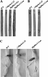
Infection assays of Mgrho3 deletion mutants. (A) Rice leaves of cultivar CO39 were sprayed with conidium suspensions from the wild-type strain (70-15), ΔMgrho3-22, MgRho3-Ect, and MgRho3-Com. Typical leaves were scanned 7 days after inoculation. (B) Wounded rice leaves of cultivar CO39 were inoculated with conidia (5 × 10⁴ conidia/ml) from strains 70-15, ΔMgrho3-22, and MgRho3-Com. Typical leaves were photographed 5 days after inoculation. (C) A root infection assay was carried out as described elsewhere (9).
FIG. 6.
FIG. 6.
Appressorium formation of the Mgrho3 deletion mutant on artificial hydrophobic surfaces. Conidia were incubated on the surface of hydrophobic GelBond films as described in Materials and Methods. Healthy appressoria from the wild-type strain (WT) and MgRho3-Com versus abnormal appressoria from the ΔMgrho3-22 strain were examined with differential interference contrast microscopy.
FIG. 7.
FIG. 7.
Penetration assays of the Mgrho3 deletion mutant on onion epidermal cells. Conidial suspensions (around 1,000 conidia in 20 μl) of wild-type strain 70-15 (WT), ΔMgrho3-22, MgRho3-Com, and MgRho3-Ect were inoculated on strips of onion epidermis as described by Xu et al. (44). Infectious hyphae were photographed 1 day after inoculation (top panel) and 2 days after inoculation (lower panel) by using differential interference contrast microscopy. A, appressorium; C, conidium; H, hypha; IF, infectious hypha.
FIG. 8.
FIG. 8.
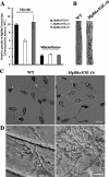
Morphology of MgRho3 overexpression mutants. (A) Total RNA samples (approximately 5 μg per reaction mixture) isolated from mycelia of M. grisea strains 70-15 (wild type [WT]) and MgRho3 overexpression transformants (MgRho3OE-8, -12, and -16) were subjected to real-time PCR. (B) Rice leaves of cultivar CO39 were sprayed with conidial suspensions from the wild-type strain (70-15) and MgRho3OE-16. Typical leaves were scanned 7 days after inoculation. (C) Appressoria examined with differential interference contrast microscopy 8 h after conidia were incubated on the surface of hydrophobic GelBond films as described in Materials and Methods. (D) Conidial suspensions (around 1,000 condia in 20 μl) of M. grisea strain 70-15 (WT) and MgRho3OE-16 were inoculated on strips of onion epidermis. Infectious hyphae were photographed 2 days after inoculation.
FIG. 9.
FIG. 9.
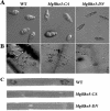
Morphology and infection assay of MgRho3 dominant mutants. (A) Conidia of 70-15 (wild type [WT]), MgRho3-CA, and MgRho3-DN cultured on oatmeal agar plates for 10 days. (B) Conidial suspensions (around 1,000 conidia in 20 μl) of M. grisea strain 70-15 (WT) and MgRho3 dominant mutants were inoculated on strips of onion epidermis. Infectious hyphae were photographed 2 days after inoculation. (C) Rice leaves of cultivar CO39 were sprayed with conidial suspensions from the wild-type strain (70-15), MgRho3-CA, and MgRho3-DN. A, appressorium; C, conidium; H, hypha; IF, infectious hypha. All were examined with differential interference contrast microscopy.

Similar articles

Cited by

References

    1. Adachi, K., and J. E. Hamer. 1998. Divergent cAMP signaling pathways regulate growth and pathogenesis in the rice blast fungus Magnaporthe grisea. Plant Cell 10:1361-1374. - PMC - PubMed
    1. Adamo, J. E., G. Rossi, and P. Brennwald. 1999. The Rho GTPase Rho3 has a direct role in exocytosis that is distinct from its role in actin polarity. Mol. Biol. Cell 10:4121-4133. - PMC - PubMed
    1. Bourett, T. M., and R. J. Howard. 1992. Actin in penetration pegs of the fungal rice blast pathogen, Magnaporthe grisea. Protoplasma 168:20-26.
    1. Bourett, T. M., J. A. Sweigard, K. J. Czymmek, A. Carroll, and R. J. Howard. 2002. Reef coral fluorescent proteins for visualizing fungal pathogens. Fungal Genet. Biol. 37:211-220. - PubMed
    1. Boyce, K. J., M. J. Hynes, and A. Andrianopoulos. 2001. The CDC42 homolog of the dimorphic fungus Penicillium marneffei is required for correct cell polarization during growth but not development. J. Bacteriol. 183:3447-3457. - PMC - PubMed

Publication types

MeSH terms

Substances

LinkOut - more resources