. 2007 Dec;293(6):H3379-87.
doi: 10.1152/ajpheart.00967.2007. Epub 2007 Oct 12.

The visceral pericardium: macromolecular structure and contribution to passive mechanical properties of the left ventricle

Affiliations

The visceral pericardium: macromolecular structure and contribution to passive mechanical properties of the left ventricle

Paul D Jöbsis et al. Am J Physiol Heart Circ Physiol. 2007 Dec.

Abstract

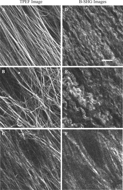
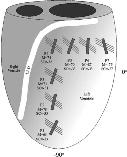
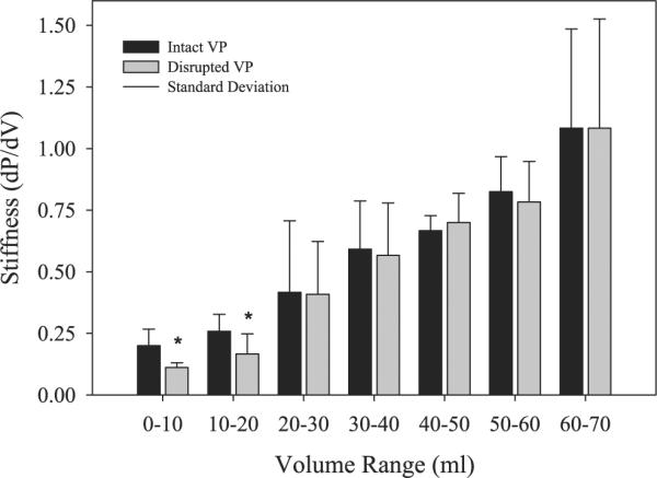
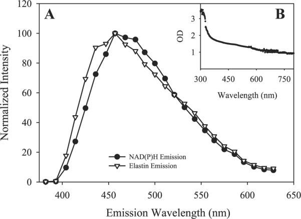
Much attention has been focused on the passive mechanical properties of the myocardium, which determines left ventricular (LV) diastolic mechanics, but the significance of the visceral pericardium (VP) has not been extensively studied. A unique en face three-dimensional volumetric view of the porcine VP was obtained using two-photon excitation fluorescence to detect elastin and backscattered second harmonic generation to detect collagen, in addition to standard light microscopy with histological staining. Below a layer of mesothelial cells, collagen and elastin fibers, extending several millimeters, form several distinct layers. The configuration of the collagen and elastin layers as well as the location of the VP at the epicardium providing a geometric advantage led to the hypothesis that VP mechanical properties play a role in the residual stress and passive stiffness of the heart. The removal of the VP by blunt dissection from porcine LV slices changed the opening angle from 53.3 +/- 10.3 to 27.3 +/- 5.7 degrees (means +/- SD, P < 0.05, n = 4). In four porcine hearts where the VP was surgically disrupted, a significant decrease in opening angle was found (35.5 +/- 4.0 degrees ) as well as a rightward shift in the ex vivo pressure-volume relationship before and after disruption and a decrease in LV passive stiffness at lower LV volumes (P < 0.05). These data demonstrate the significant and previously unreported role that the VP plays in the residual stress and passive stiffness of the heart. Alterations in this layer may occur in various disease states that effect diastolic function.


Figures

Fig. 1
Fig. 1
Micrographs of cardiac tissue with Movat staining. Collagen appears gold, and elastin fibers appear dark purple or black. The intact visceral pericardium (VP; A) appears to consist primarily of collagen with distinct layers of elastin fibers. Following blunt dissection, the VP is almost completely removed (B). The intact endocardial membrane (C) appears to be rich in elastin relative to the collagen density. The 50-μm scale bar in A applies to all images.
Fig. 2
Fig. 2
Two-photon excitation fluorescence (TPEF) and backscattered second harmonic generation (B-SHG) micrographs of a relatively thin area of VP collected simultaneously using a Zeiss LSM 510 META microscope. At the surface (8 μm) of the VP, parallel elastin rods (A) run along the long axis of collagen fibers (D). In the midsection of the VP (16 μm), elastin rods (B) form a network that appears to be independent of collagen orientation (E). At the VP and myocyte interface (30 μm), elastin rods (C) again appear to follow the orientation of collagen fibers (F). This pattern was found in most VP samples imaged in this manner, although the pattern was interrupted by blood vessels within the VP. The scale bar in D represents 20 μm and applies to all images.
Fig. 3
Fig. 3
TPEF and B-SHG micrographs of a thin section of endocardial membrane. Images show the endocardial membrane at depths of 4 μm (A and D), 15 μm (B and E), and 30 μm (C and F). The endocardial membrane has a similar pattern of collagen and elastin, but the elastin and collagen appear thinner and more diffuse and do not follow the patterns found in the VP. Imaging parameters, laser power, and filter settings were identical to those used in Fig. 2. The scale bar in D represents 20 μm and applies to all images.
Fig. 4
Fig. 4
Diagram of the left ventricle (LV) and right ventricle showing the imaging locations [positions 17 (P1–P7)] for the fiber orientation survey. LAD, left anterior descending coronary artery. Surface collagen and elastin (SC) of the VP run parallel to each other and are represented by the long narrow lines. The shorter and thicker solid lines represent the average myocyte (M) orientation from the VP-myocardium interface. The surface collagen and elastin orientation and VP-myocardium interface myocyte orientation (means ± SD, n = 4) are as follows: P1, −34 ± 9° and 63 ± 3°; P2, −35 ± 21° and 78 ± 3°; P3, −31 ± 8° and 71 ± 1°; P4, −36 ± 14° and 74 ± 9°; P5, −38 ± 12° and 70 ± 23°; P6, −34 ± 12° and 87 ± 43°; and P7, −27 ± 10° and −75 ± 7°.
Fig. 5
Fig. 5
Representative photographs of the opening angle of LV slices with intact (A), peeled (B), and disrupted VPs (C), respectively. ANOVA and Tukey analysis of the opening angles showed that the average opening angle (n = 4) for LV slices with either peeled (27.4 ± 5.7°) or disrupted VP (35.5 ± 4.0°) had significantly (P < 0.05) reduced opening angles compared with LV slices with an intact VP (53.3 ± 10.3°).
Fig. 6
Fig. 6
Pressure-volume relationship of the LV before and after VP disruption. Disruption of the VP caused a highly significant shift in the entire pressure-volume relationship (P < 0.05 by paired t-test, n = 4).
Fig. 7
Fig. 7
LV passive stiffness [change in pressure over volume (dP/dV)] before (intact VP) and after VP disruption. VP disruption significantly reduced LV stiffness at lower volumes, as indicated by the asterisks (0–20 ml, P < 0.05), but not in higher volumes (20–70 ml, P < 0.05). This suggests that the mechanical effect of VP disruption is primarily due to disruption of the elastin elements of the VP and that other elements remain viable at the higher ranges measured in this study.
Fig. 8
Fig. 8
Elastin fiber length change during systole versus fiber angle. Two estimates are shown for no appreciable circumferential shortening (Ecc)on the epicardial surface or 2% Ecc. The range of measured elastin fiber angles fell in the region where stretching occurred during systole, whereas the range of measured myofiber angles resulted in the expected 10% shortening. Stretching of elastin fibers is an effective way to store mechanical energy to aid in LV relaxation and untwisting at the onset of diastole.
Fig. 9
Fig. 9
A: elastin and NAD(P)H fluorescence collected using the META detector of the Zeiss LSM 510 microscope. The similar absorbance spectra caused difficulty in optically separating the emissions. In B, the scattering spectrum of the VP layer, once it had been removed from the myocardium, showed a high degree of scattering from an ~100-μm-thick layer.

References

    1. Agostoni E, Bodega F, Zocchi L. Equivalent radius of paracellular “pores” of the mesothelium. J Appl Physiol. 1999;87:538–544. - PubMed
    1. Aletras AH, Balaban RS, Wen H. High-resolution strain analysis of the human heart with fast-DENSE. J Magn Reson. 1999;140:41–57. - PubMed
    1. Aletras AH, Wen H. Mixed echo train acquisition displacement encoding with stimulated echoes: an optimized DENSE method for in vivo functional imaging of the human heart. Magn Reson Med. 2001;46:523–534. - PMC - PubMed
    1. Bell SP, Nyland L, Tischler MD, McNabb M, Granzier H, LeWinter MM. Alterations in the determinants of diastolic suction during pacing tachycardia. Circ Res. 2000;87:235–240. - PubMed
    1. Burlew BS, Weber KT. Connective tissue and the heart. Functional significance and regulatory mechanisms. Cardiol Clin. 2000;18:435–442. - PubMed