

Characterization of HLA class I altered phenotypes in a panel of human melanoma cell lines
- PMID: 17934731
- PMCID: PMC11030649
- DOI: 10.1007/s00262-007-0411-3
Characterization of HLA class I altered phenotypes in a panel of human melanoma cell lines
Abstract
Background: Altered HLA class I cell surface expression is one of the major mechanisms by which tumor cells escape from T lymphocytes. Immunohistochemistry-defined phenotypes of lost HLA class I expression have been described in human solid tumors, nut less information is available on melanoma cell lines.
Objectives: To describe the frequency and distribution of different types of HLA class I antigen alterations in 91 melanoma cell lines from the European Searchable Tumour Cell and Databank (ESTDAB).
Methods: The HLA class I expression was assessed by flow cytometry and HLA genotyping.
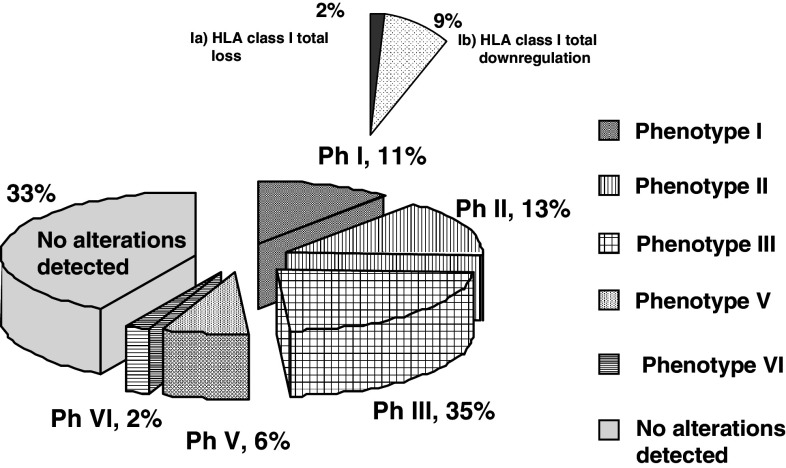
Results: We found various types of HLA class I cell surface alterations in about 67% of the melanoma cell lines. These alterations range from total to selective HLA class I loss due to loss of heterozygosity (LOH), haplotype loss, beta2-microglobulin gene mutation, and/or total or selective down-regulation of HLA class I molecules. The most frequently observed phenotype is down-regulation of HLA-B locus that was reversible after treatment with IFN -gamma.
Conclusions: In general, HLA class I alterations in the majority of the cells analyzed were of regulatory nature and could be restored by IFN-gamma. Analysis of the frequency of distinct HLA class I altered phenotypes in these melanoma cell lines revealed specific differences compared to other types of tumors.
Figures
Similar articles
-
HLA and melanoma: multiple alterations in HLA class I and II expression in human melanoma cell lines from ESTDAB cell bank.Cancer Immunol Immunother. 2009 Sep;58(9):1507-15. doi: 10.1007/s00262-009-0701-z. Epub 2009 Apr 2. Cancer Immunol Immunother. 2009. PMID: 19340423 Free PMC article. Review.
-
Immune escape of cancer cells with beta2-microglobulin loss over the course of metastatic melanoma.Int J Cancer. 2014 Jan 1;134(1):102-13. doi: 10.1002/ijc.28338. Epub 2013 Jul 16. Int J Cancer. 2014. PMID: 23784959
-
High frequency of homozygosity of the HLA region in melanoma cell lines reveals a pattern compatible with extensive loss of heterozygosity.Cancer Immunol Immunother. 2005 Feb;54(2):141-8. doi: 10.1007/s00262-004-0561-5. Epub 2004 Oct 1. Cancer Immunol Immunother. 2005. PMID: 15592718 Free PMC article.
-
The coincidence of chromosome 15 aberrations and beta2-microglobulin gene mutations is causative for the total loss of human leukocyte antigen class I expression in melanoma.Clin Cancer Res. 2006 Jun 1;12(11 Pt 1):3297-305. doi: 10.1158/1078-0432.CCR-05-2174. Clin Cancer Res. 2006. PMID: 16740750
-
Beta2-microglobulin gene mutations in human melanoma cells: molecular characterization and implications for immune surveillance.Melanoma Res. 1997 Aug;7 Suppl 2:S67-74. Melanoma Res. 1997. PMID: 9578419 Review.
Cited by
-
Genome-wide association study identifies 14 novel risk alleles associated with basal cell carcinoma.Nat Commun. 2016 Aug 19;7:12510. doi: 10.1038/ncomms12510. Nat Commun. 2016. PMID: 27539887 Free PMC article.
-
The immune-related role of beta-2-microglobulin in melanoma.Front Oncol. 2022 Aug 16;12:944722. doi: 10.3389/fonc.2022.944722. eCollection 2022. Front Oncol. 2022. PMID: 36046045 Free PMC article. Review.
-
Analysis of HLA-ABC locus-specific transcription in normal tissues.Immunogenetics. 2010 Dec;62(11-12):711-9. doi: 10.1007/s00251-010-0470-z. Epub 2010 Sep 15. Immunogenetics. 2010. PMID: 20842357
-
HLA and melanoma: multiple alterations in HLA class I and II expression in human melanoma cell lines from ESTDAB cell bank.Cancer Immunol Immunother. 2009 Sep;58(9):1507-15. doi: 10.1007/s00262-009-0701-z. Epub 2009 Apr 2. Cancer Immunol Immunother. 2009. PMID: 19340423 Free PMC article. Review.
-
Adaptive Resistance to Cancer Immunotherapy.Adv Exp Med Biol. 2017;1036:213-227. doi: 10.1007/978-3-319-67577-0_14. Adv Exp Med Biol. 2017. PMID: 29275474 Review.
References
-
- Benitez R, Godelaine D, Lopez-Nevot MA, Brasseur F, Jimenez P, Marchand M, Oliva MR, van Baren N, Cabrera T, Andry G, Landry C, Ruiz-Cabello F, Boon T, Garrido F. Mutations of the beta2-microglobulin gene result in a lack of HLA class I molecules on melanoma cells of two patients immunized with MAGE peptides. Tissue Antigens. 1998;52:520. doi: 10.1111/j.1399-0039.1998.tb03082.x. - DOI - PubMed
-
- Cabrera CM, Jimenez P, Cabrera T, Esparza C, Ruiz-Cabello F, Garrido F. Total loss of MHC class I in colorectal tumors can be explained by two molecular pathways: beta2-microglobulin inactivation in MSI-positive tumors and LMP7/TAP2 downregulation in MSI-negative tumors. Tissue Antigens. 2003;61:211. doi: 10.1034/j.1399-0039.2003.00020.x. - DOI - PubMed
Publication types
MeSH terms
Substances
LinkOut - more resources
Full Text Sources
Medical
Research Materials
