

VEGF-C alters barrier function of cultured lymphatic endothelial cells through a VEGFR-3-dependent mechanism
- PMID: 17935478
- PMCID: PMC3001341
- DOI: 10.1089/lrb.2007.1004
VEGF-C alters barrier function of cultured lymphatic endothelial cells through a VEGFR-3-dependent mechanism
Abstract
Background: The lymphatic endothelium is an important semi-permeable barrier separating lymph from the interstitial space. However, there is currently a limited understanding of the lymphatic endothelial barrier and the mechanisms of lymph formation. The objectives of this study were to investigate the potential active role of lymphatic endothelial cells in barrier regulation, and to test whether the endothelial cell agonists VEGF-A and VEGF-C can alter lymphatic endothelial barrier function.
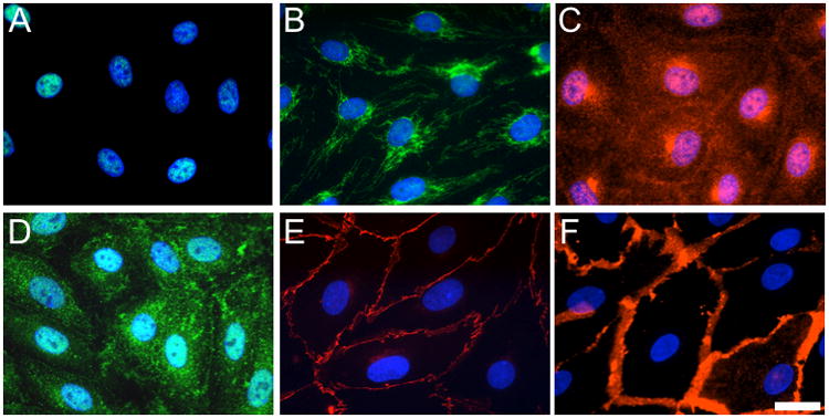
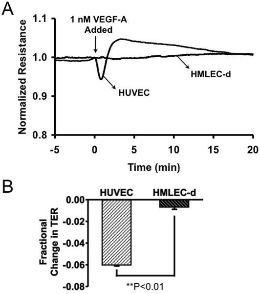
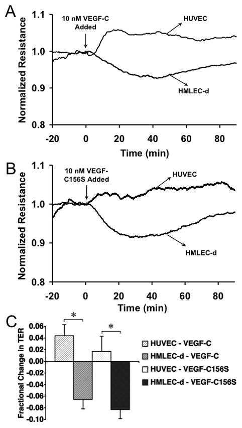
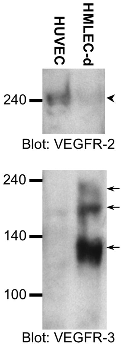
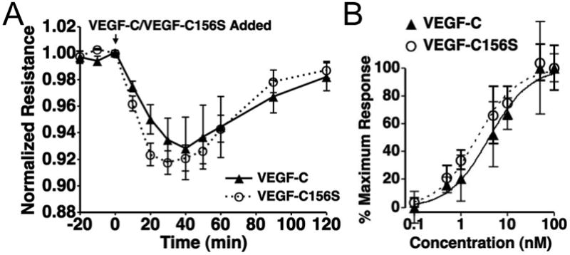
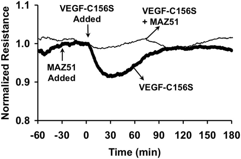
Methods and results: Cultured adult human dermal microlymphatic endothelial cells (HMLEC-d) and human umbilical vein endothelial cells (HUVEC) were respectively used as models of lymphatic and vascular endothelium. Transendothelial electrical resistance (TER) of endothelial monolayers served as an index of barrier function. Cells were treated with VEGF-A, VEGF-C, or the VEGFR-3 selective mutant VEGF-C156S. MAZ51 was used to inhibit VEGFR-3 signaling. The results show that while VEGF-A causes a time-dependent decrease in TER in HUVEC, there is no response in HMLEC-d. In contrast, VEGF-C and VEGF-C156S cause a similar decrease in TER in HMLEC-d that is not observed in HUVEC. These results corresponded to the protein expression of VEGFR-2 and VEGFR-3 in these cell types, determined by Western blotting. In addition, the VEGF-C- and VEGF-C156S-induced TER changes were inhibited by MAZ51.
Conclusions: The results indicate differential responses of the lymphatic and vascular endothelial barriers to VEGF-A and VEGF-C. Furthermore, our data suggest that VEGF-C alters lymphatic endothelial function through a mechanism involving VEGFR-3.
Figures
Comment in
-
Molecular insights into the microvascular regulation of lymph formation.Lymphat Res Biol. 2007;5(3):149-50. doi: 10.1089/lrb.2007.5301. Lymphat Res Biol. 2007. PMID: 18035932 No abstract available.
Similar articles
-
Lymphatic reprogramming of microvascular endothelial cells by CEA-related cell adhesion molecule-1 via interaction with VEGFR-3 and Prox1.Blood. 2007 Dec 15;110(13):4223-33. doi: 10.1182/blood-2007-06-097592. Epub 2007 Aug 30. Blood. 2007. PMID: 17761831
-
The tyrosine kinase inhibitor cediranib blocks ligand-induced vascular endothelial growth factor receptor-3 activity and lymphangiogenesis.Cancer Res. 2008 Jun 15;68(12):4754-62. doi: 10.1158/0008-5472.CAN-07-5809. Cancer Res. 2008. PMID: 18559522
-
Suppression of lymphangiogenesis in human lymphatic endothelial cells by simultaneously blocking VEGF-C and VEGF-D/VEGFR-3 with norcantharidin.Int J Oncol. 2012 Nov;41(5):1762-72. doi: 10.3892/ijo.2012.1603. Epub 2012 Aug 23. Int J Oncol. 2012. PMID: 22922710
-
[Characterization of markers and growth factors for lymphatic endothelium].Postepy Biochem. 2005;51(2):209-14. Postepy Biochem. 2005. PMID: 16209358 Review. Polish.
-
The role of the VEGF-C/VEGFR-3 axis in cancer progression.Br J Cancer. 2007 Feb 26;96(4):541-5. doi: 10.1038/sj.bjc.6603487. Epub 2006 Dec 12. Br J Cancer. 2007. PMID: 17164762 Free PMC article. Review.
Cited by
-
A 3D Human Lymphatic Vessel-on-Chip Reveals the Roles of Interstitial Flow and VEGF-A/C for Lymphatic Sprouting and Discontinuous Junction Formation.Cell Mol Bioeng. 2023 Aug 24;16(4):325-339. doi: 10.1007/s12195-023-00780-0. eCollection 2023 Aug. Cell Mol Bioeng. 2023. PMID: 37811004 Free PMC article.
-
PEG-Immobilized Keratin for Protein Drug Sequestration and pH-Mediated Delivery.J Drug Deliv. 2016;2016:7843951. doi: 10.1155/2016/7843951. Epub 2016 Jan 20. J Drug Deliv. 2016. PMID: 26904294 Free PMC article.
-
Lymphatic-specific intracellular modulation of receptor tyrosine kinase signaling improves lymphatic growth and function.Sci Signal. 2021 Aug 10;14(695):eabc0836. doi: 10.1126/scisignal.abc0836. Sci Signal. 2021. PMID: 34376570 Free PMC article.
-
Lymphatic endothelial cells adapt their barrier function in response to changes in shear stress.Lymphat Res Biol. 2009 Dec;7(4):229-37. doi: 10.1089/lrb.2009.0015. Lymphat Res Biol. 2009. PMID: 20143922 Free PMC article.
-
Acute alcohol intoxication-induced microvascular leakage.Alcohol Clin Exp Res. 2014 Sep;38(9):2414-26. doi: 10.1111/acer.12525. Alcohol Clin Exp Res. 2014. PMID: 25257290 Free PMC article.
References
-
- Adair TH, Guyton AC. Lymph formation and its modification in the lymphatic system. In: Johnston MG, editor. Experimental Biology of the Lymphatic Circulation. New York: Elsevier; 1985. pp. 13–44.
-
- Schmid-Schonbein GW. The second valve system in lymphatics. Lymphat Res Biol. 2003;1:25–9. discussion 9–31. - PubMed
-
- Mendoza E, Schmid-Schonbein GW. A model for mechanics of primary lymphatic valves. J Biomech Eng. 2003;125:407–14. - PubMed
-
- Trzewik J, Mallipattu SK, Artmann GM, Delano FA, Schmid-Schonbein GW. Evidence for a second valve system in lymphatics: endothelial microvalves. Faseb J. 2001;15:1711–7. - PubMed
-
- Yuan SY. Signal transduction pathways in enhanced microvascular permeability. Microcirculation. 2000;7:395–403. - PubMed
Publication types
MeSH terms
Substances
Grants and funding
LinkOut - more resources
Full Text Sources
Research Materials
Miscellaneous
