

SOX9 transduction of a human chondrocytic cell line identifies novel genes regulated in primary human chondrocytes and in osteoarthritis
- PMID: 17935617
- PMCID: PMC2212576
- DOI: 10.1186/ar2311
SOX9 transduction of a human chondrocytic cell line identifies novel genes regulated in primary human chondrocytes and in osteoarthritis
Abstract
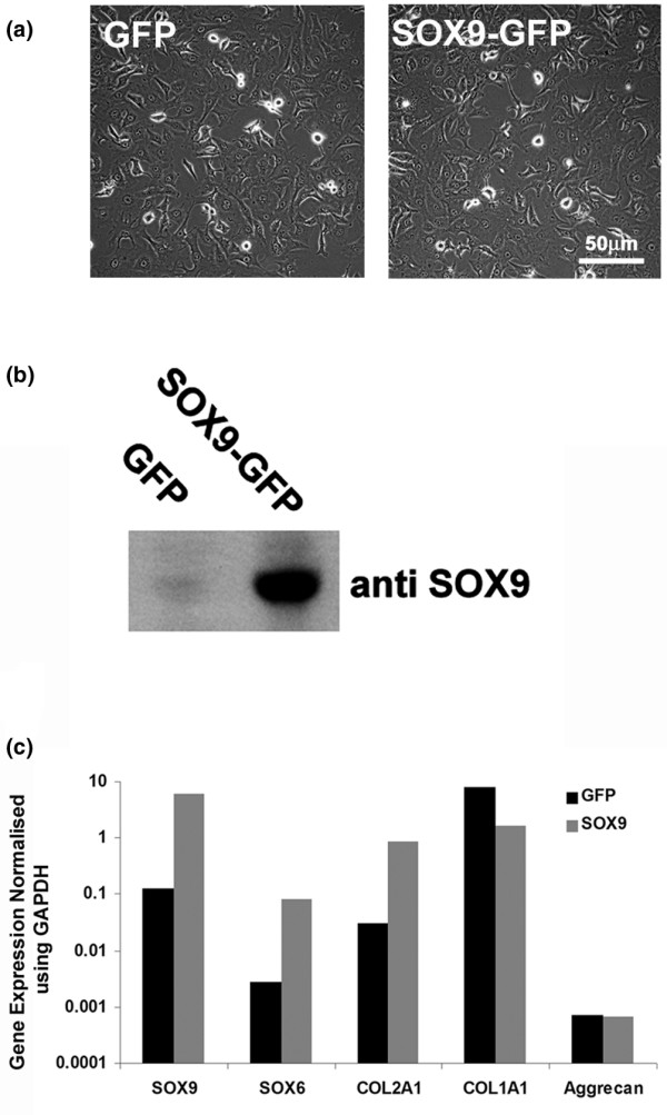
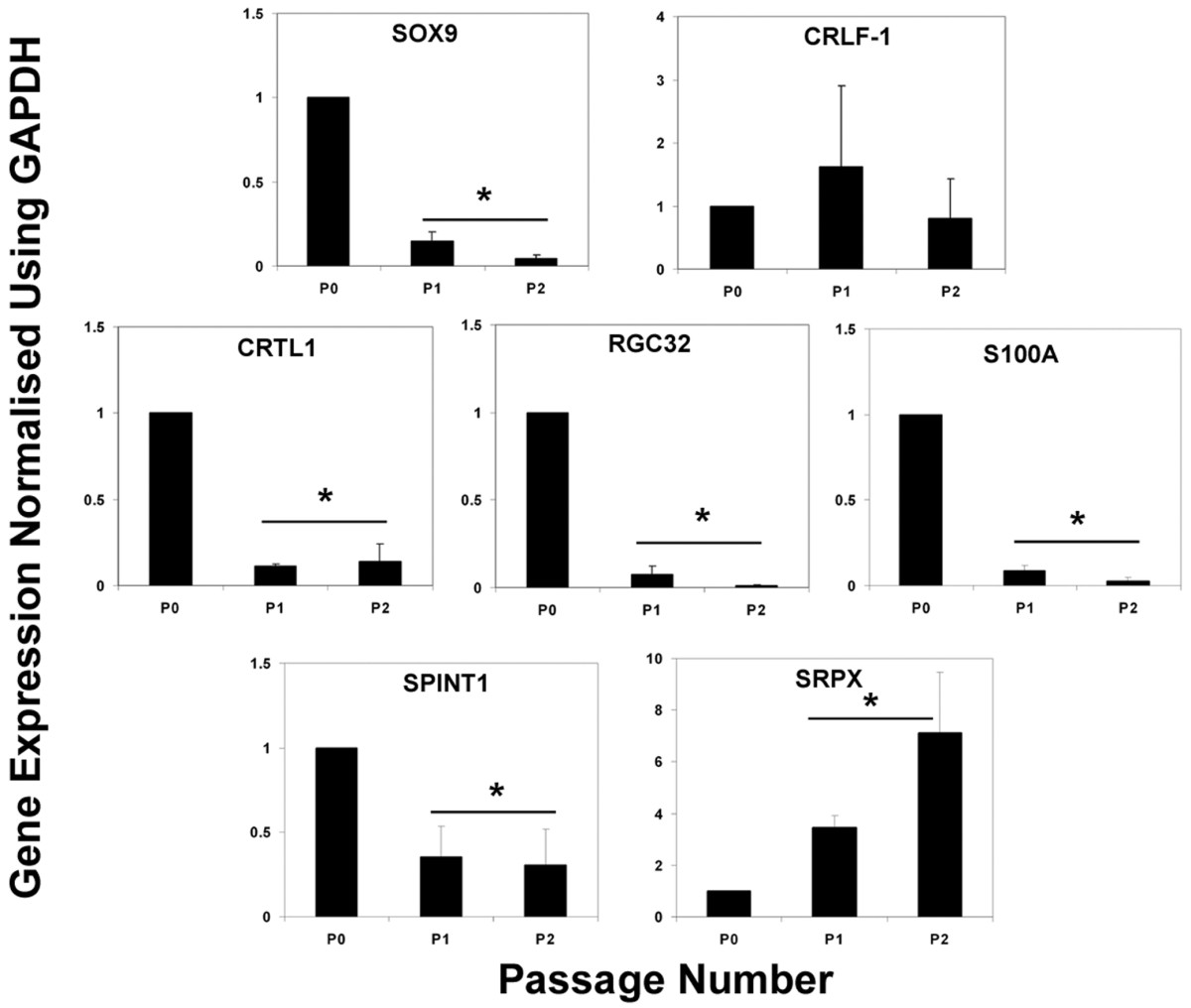
The transcription factor SOX9 is important in maintaining the chondrocyte phenotype. To identify novel genes regulated by SOX9 we investigated changes in gene expression by microarray analysis following retroviral transduction with SOX9 of a human chondrocytic cell line (SW1353). From the results the expression of a group of genes (SRPX, S100A1, APOD, RGC32, CRTL1, MYBPH, CRLF1 and SPINT1) was evaluated further in human articular chondrocytes (HACs). First, the same genes were investigated in primary cultures of HACs following SOX9 transduction, and four were found to be similarly regulated (SRPX, APOD, CRTL1 and S100A1). Second, during dedifferentiation of HACs by passage in monolayer cell culture, during which the expression of SOX9 progressively decreased, four of the genes (S100A1, RGC32, CRTL1 and SPINT1) also decreased in their expression. Third, in samples of osteoarthritic (OA) cartilage, which had decreased SOX9 expression compared with age-matched controls, there was decreased expression of SRPX, APOD, RGC32, CRTL1 and SPINT1. The results showed that a group of genes identified as being upregulated by SOX9 in the initial SW1353 screen were also regulated in expression in healthy and OA cartilage. Other genes initially identified were differently expressed in isolated OA chondrocytes and their expression was unrelated to changes in SOX9. The results thus identified some genes whose expression appeared to be linked to SOX9 expression in isolated chondrocytes and were also altered during cartilage degeneration in osteoarthritis.
Figures

Similar articles
-
Retroviral transduction with SOX9 enhances re-expression of the chondrocyte phenotype in passaged osteoarthritic human articular chondrocytes.Osteoarthritis Cartilage. 2005 Jan;13(1):80-9. doi: 10.1016/j.joca.2004.10.011. Osteoarthritis Cartilage. 2005. PMID: 15639641
-
Transduction of passaged human articular chondrocytes with adenoviral, retroviral, and lentiviral vectors and the effects of enhanced expression of SOX9.Tissue Eng. 2004 Mar-Apr;10(3-4):575-84. doi: 10.1089/107632704323061933. Tissue Eng. 2004. PMID: 15165474
-
Transforming growth factor alpha suppression of articular chondrocyte phenotype and Sox9 expression in a rat model of osteoarthritis.Arthritis Rheum. 2007 Nov;56(11):3693-705. doi: 10.1002/art.22968. Arthritis Rheum. 2007. PMID: 17968906
-
SOX9 expression does not correlate with type II collagen expression in adult articular chondrocytes.Matrix Biol. 2003 Jun;22(4):363-72. doi: 10.1016/s0945-053x(03)00049-0. Matrix Biol. 2003. PMID: 12935820
-
Transcriptional mechanisms of chondrocyte differentiation.Matrix Biol. 2000 Sep;19(5):389-94. doi: 10.1016/s0945-053x(00)00094-9. Matrix Biol. 2000. PMID: 10980415 Review.
Cited by
-
Transcription Factors in Cartilage Homeostasis and Osteoarthritis.Biology (Basel). 2020 Sep 14;9(9):290. doi: 10.3390/biology9090290. Biology (Basel). 2020. PMID: 32937960 Free PMC article. Review.
-
Suppression of CRLF1 promotes the chondrogenic differentiation of bone marrow-derived mesenchymal stem and protects cartilage tissue from damage in osteoarthritis via activation of miR-320.Mol Med. 2021 Sep 22;27(1):116. doi: 10.1186/s10020-021-00369-1. Mol Med. 2021. PMID: 34551709 Free PMC article.
-
Exploring the role of directly coupled alternating electric fields on chondrocyte morphology and redifferentiation capacity with a focus on sex differences.J Exp Orthop. 2025 May 6;12(2):e70261. doi: 10.1002/jeo2.70261. eCollection 2025 Apr. J Exp Orthop. 2025. PMID: 40330809 Free PMC article.
-
A genome-wide association study in Han Chinese identifies new susceptibility loci for ankylosing spondylitis.Nat Genet. 2011 Dec 4;44(1):73-7. doi: 10.1038/ng.1005. Nat Genet. 2011. PMID: 22138694
-
Hyperosmolarity regulates SOX9 mRNA posttranscriptionally in human articular chondrocytes.Am J Physiol Cell Physiol. 2009 Oct;297(4):C898-906. doi: 10.1152/ajpcell.00571.2008. Epub 2009 Aug 5. Am J Physiol Cell Physiol. 2009. PMID: 19657054 Free PMC article.
References
-
- Brew CJ, Andrew JG, Boot-Handford R, Hardingham TE. Late osteoarthritic cartilage shows down regulation of SOX9 and aggrecan expression but little evidence of chondrocyte hypertrophy. Trans Orthop Res Soc. 2004;50:938.
Publication types
MeSH terms
Substances
Grants and funding
LinkOut - more resources
Full Text Sources
Other Literature Sources
Medical
Research Materials
Miscellaneous
